Mass: 18.015 Da / Num. of mol.: 154 / Source method: isolated from a natural source / Formula: H2O
-
Experimental details
-
Experiment
Experiment
Method: X-RAY DIFFRACTION / Number of used crystals: 1
-
Sample preparation
Crystal
Density Matthews: 2.42 Å3/Da / Density % sol: 49.26 %
Crystal grow
Temperature: 298 K / Method: vapor diffusion, sitting drop / pH: 7.5 Details: WELL SOLUTION: 25% PEG 3350, 0.1 M NH4 SULPHATE, 0.1 M HEPES PH 7.5. PROTEIN SOLUTION: 15 MG/ML. DROPS: 1:1 RATIO OF WELL:PROTEIN SOLUTION. CRYSTALS CRYOPROTECTED BY TRANSFER TO DROP ...Details: WELL SOLUTION: 25% PEG 3350, 0.1 M NH4 SULPHATE, 0.1 M HEPES PH 7.5. PROTEIN SOLUTION: 15 MG/ML. DROPS: 1:1 RATIO OF WELL:PROTEIN SOLUTION. CRYSTALS CRYOPROTECTED BY TRANSFER TO DROP CONTAINING MOTHER LIQUOR TO WHICH MPD WAS ADDED TO 25%, , pH 7.50, VAPOR DIFFUSION, SITTING DROP, temperature 298.0K
Resolution: 2→23.69 Å / Cor.coef. Fo:Fc: 0.955 / Cor.coef. Fo:Fc free: 0.931 / SU B: 8.289 / SU ML: 0.119 / Cross valid method: THROUGHOUT / ESU R: 0.179 / ESU R Free: 0.161 / Stereochemistry target values: MAXIMUM LIKELIHOOD Details: HYDROGENS HAVE BEEN ADDED IN THE RIDING POSITIONS. ATOM RECORD CONTAINS SUM OF TLS AND RESIDUAL B FACTORS, ANISOU RECORD CONTAINS SUM OF TLS AND RESIDUAL U FACTORS.
Rfactor
Num. reflection
% reflection
Selection details
Rfree
0.2316
1062
4.9 %
RANDOM
Rwork
0.18917
-
-
-
obs
0.19118
20747
95.3 %
-
all
-
21809
-
-
Solvent computation
Ion probe radii: 0.8 Å / Shrinkage radii: 0.8 Å / VDW probe radii: 1.2 Å / Solvent model: BABINET MODEL WITH MASK
Displacement parameters
Biso mean: 27.811 Å2
Baniso -1
Baniso -2
Baniso -3
1-
1.06 Å2
0.53 Å2
0 Å2
2-
-
1.06 Å2
0 Å2
3-
-
-
-1.59 Å2
Refinement step
Cycle: LAST / Resolution: 2→23.69 Å
Protein
Nucleic acid
Ligand
Solvent
Total
Num. atoms
2172
0
30
156
2358
Refine LS restraints
Refine-ID
Type
Dev ideal
Dev ideal target
Number
X-RAY DIFFRACTION
r_bond_refined_d
0.011
0.022
2254
X-RAY DIFFRACTION
r_angle_refined_deg
1.156
1.963
3057
X-RAY DIFFRACTION
r_dihedral_angle_1_deg
5.13
5
278
X-RAY DIFFRACTION
r_dihedral_angle_2_deg
32.222
23.529
102
X-RAY DIFFRACTION
r_dihedral_angle_3_deg
12.401
15
372
X-RAY DIFFRACTION
r_dihedral_angle_4_deg
20.531
15
17
X-RAY DIFFRACTION
r_chiral_restr
0.079
0.2
328
X-RAY DIFFRACTION
r_gen_planes_refined
0.004
0.02
1733
X-RAY DIFFRACTION
r_nbd_refined
0.195
0.2
1067
X-RAY DIFFRACTION
r_nbtor_refined
0.3
0.2
1562
X-RAY DIFFRACTION
r_xyhbond_nbd_refined
0.121
0.2
144
X-RAY DIFFRACTION
r_symmetry_vdw_refined
0.155
0.2
41
X-RAY DIFFRACTION
r_symmetry_hbond_refined
0.107
0.2
14
X-RAY DIFFRACTION
r_mcbond_it
0.599
1.5
1433
X-RAY DIFFRACTION
r_mcangle_it
0.939
2
2223
X-RAY DIFFRACTION
r_scbond_it
1.451
3
961
X-RAY DIFFRACTION
r_scangle_it
2.329
4.5
834
LS refinement shell
Resolution: 2→2.052 Å / Total num. of bins used: 20
Rfactor
Num. reflection
% reflection
Rfree
0.242
93
-
Rwork
0.212
1549
-
obs
-
-
97.8 %
Refinement TLS params.
Method: refined / Refine-ID: X-RAY DIFFRACTION
ID
L11 (°2)
L12 (°2)
L13 (°2)
L22 (°2)
L23 (°2)
L33 (°2)
S11 (Å °)
S12 (Å °)
S13 (Å °)
S21 (Å °)
S22 (Å °)
S23 (Å °)
S31 (Å °)
S32 (Å °)
S33 (Å °)
T11 (Å2)
T12 (Å2)
T13 (Å2)
T22 (Å2)
T23 (Å2)
T33 (Å2)
Origin x (Å)
Origin y (Å)
Origin z (Å)
1
52.3212
-10.2619
3.0468
16.7594
-4.1977
1.2027
-1.3328
-3.8486
-0.8284
1.6378
1.4336
2.3378
-0.8689
-0.259
-0.1008
0.5822
0.2976
0.2799
0.398
-0.0178
0.3728
-23.5049
15.895
6.7817
2
5.6051
-4.3126
6.4159
15.8154
-12.0783
29.9614
-0.0209
-0.1316
0.1512
1.0372
0.2688
-0.1928
-0.1092
0.3311
-0.2479
0.2177
0.0751
0.0634
-0.0546
-0.0442
0.1554
-24.5506
23.3388
-2.2199
3
3.5596
-1.44
-1.7272
15.3352
3.3101
11.4513
-0.0798
0.2148
-0.2242
0.3223
-0.0615
0.4156
0.8852
-0.1297
0.1413
0.2131
0.0652
-0.0064
0.0183
0.0334
0.1242
-26.3481
16.8901
-19.0731
4
6.3916
-2.6059
0.8989
1.082
-0.0721
4.5529
-0.1467
-0.2665
-0.1607
0.1439
-0.0111
-0.0156
0.3753
-0.3753
0.1579
0.358
0.0244
0.0712
0.0023
-0.0142
0.1223
-24.1384
15.7756
-8.054
5
0.9278
-1.0216
0.0987
1.7382
0.0568
0.0551
0.1714
0.1004
0.0465
-0.1808
-0.0892
-0.0147
-0.1583
-0.0306
-0.0822
0.2173
0.0149
0.031
0.1042
0.0066
0.1338
-12.5487
7.8593
-11.7383
6
0.9816
0.3196
-0.6064
2.3233
0.268
2.4454
0.066
0.0866
-0.0768
0.0556
-0.0729
0.0815
0.1043
-0.2141
0.0069
0.1343
0.0006
0.0059
0.1158
-0.0028
0.1558
-11.9762
-5.2213
-4.536
7
1.3403
0.0946
-0.1955
3.8604
0.6872
3.6522
-0.0602
-0.0963
-0.114
0.2218
0.0203
-0.259
0.2027
0.2173
0.04
0.1136
0.0216
-0.0191
0.0881
-0.0076
0.1843
-2.4782
-7.6459
-2.1622
8
15.6746
-2.6792
16.0375
96.0936
-32.015
25.3694
-2.0645
-2.919
-1.9897
2.7216
4.9309
-4.0892
-1.1194
-0.8358
-2.8664
0.5296
0.0908
0.15
0.6858
-0.1846
0.4681
11.407
-0.3139
-12.0321
Refinement TLS group
ID
Refine-ID
Refine TLS-ID
Auth asym-ID
Label asym-ID
Auth seq-ID
Label seq-ID
1
X-RAY DIFFRACTION
1
A
A
607 - 618
19 - 30
2
X-RAY DIFFRACTION
2
A
A
619 - 628
31 - 40
3
X-RAY DIFFRACTION
3
A
A
629 - 654
41 - 66
4
X-RAY DIFFRACTION
4
A
A
655 - 696
67 - 108
5
X-RAY DIFFRACTION
5
A
A
697 - 770
109 - 182
6
X-RAY DIFFRACTION
6
A
A
771 - 841
183 - 253
7
X-RAY DIFFRACTION
7
A
A
842 - 893
254 - 305
8
X-RAY DIFFRACTION
8
A
A
894 - 902
306 - 314
+
About Yorodumi
-
News
-
Feb 9, 2022. New format data for meta-information of EMDB entries
New format data for meta-information of EMDB entries
Version 3 of the EMDB header file is now the official format.
The previous official version 1.9 will be removed from the archive.
In the structure databanks used in Yorodumi, some data are registered as the other names, "COVID-19 virus" and "2019-nCoV". Here are the details of the virus and the list of structure data.
Jan 31, 2019. EMDB accession codes are about to change! (news from PDBe EMDB page)
EMDB accession codes are about to change! (news from PDBe EMDB page)
The allocation of 4 digits for EMDB accession codes will soon come to an end. Whilst these codes will remain in use, new EMDB accession codes will include an additional digit and will expand incrementally as the available range of codes is exhausted. The current 4-digit format prefixed with “EMD-” (i.e. EMD-XXXX) will advance to a 5-digit format (i.e. EMD-XXXXX), and so on. It is currently estimated that the 4-digit codes will be depleted around Spring 2019, at which point the 5-digit format will come into force.
The EM Navigator/Yorodumi systems omit the EMD- prefix.
Related info.:Q: What is EMD? / ID/Accession-code notation in Yorodumi/EM Navigator
Yorodumi is a browser for structure data from EMDB, PDB, SASBDB, etc.
This page is also the successor to EM Navigator detail page, and also detail information page/front-end page for Omokage search.
The word "yorodu" (or yorozu) is an old Japanese word meaning "ten thousand". "mi" (miru) is to see.
Related info.:EMDB / PDB / SASBDB / Comparison of 3 databanks / Yorodumi Search / Aug 31, 2016. New EM Navigator & Yorodumi / Yorodumi Papers / Jmol/JSmol / Function and homology information / Changes in new EM Navigator and Yorodumi
 Movie
Movie Controller

 Yorodumi
 Open data
 Basic information
 Components
 Keywords
 Function and homology information
 Homo sapiens (human)
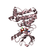
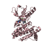
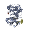
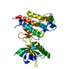
 X-RAY DIFFRACTION /
  Authors
 Citation
 Structure visualization
 Downloads & links
 Other downloads

 PDBj
PDBj


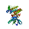
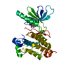
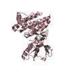
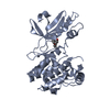
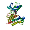
 Assembly
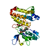


 Mass: 414.380 Da / Num. of mol.: 1 / Source method: obtained synthetically / Formula: C21H17F3N4O2
 Mass: 18.015 Da / Num. of mol.: 154 / Source method: isolated from a natural source / Formula: H2O
 Sample preparation
 Processing