 Movie
Movie Controller

[English] 日本語
 Yorodumi
- PDB-1vr2: HUMAN VASCULAR ENDOTHELIAL GROWTH FACTOR RECEPTOR 2 (KDR) KINASE ... -
+ Open data

- Basic information
| Entry | Database: PDB / ID: 1vr2 | ||||||
|---|---|---|---|---|---|---|---|
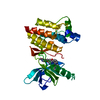
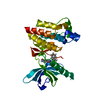
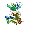
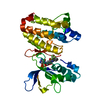
| Title | HUMAN VASCULAR ENDOTHELIAL GROWTH FACTOR RECEPTOR 2 (KDR) KINASE DOMAIN | ||||||
 Components Components | PROTEIN (VASCULAR ENDOTHELIAL GROWTH FACTOR RECEPTOR KINASE) | ||||||
 Keywords Keywords | TRANSFERASE / TYROSINE KINASE | ||||||
| Function / homology |  Function and homology information Function and homology informationpositive regulation of nitric oxide-cGMP mediated signal transduction / blood vessel endothelial cell differentiation / regulation of bone development / cellular response to hydrogen sulfide / Signaling by membrane-tethered fusions of PDGFRA or PDGFRB / Neuropilin interactions with VEGF and VEGFR / vascular endothelial growth factor binding / VEGF binds to VEGFR leading to receptor dimerization / vascular endothelial growth factor receptor-2 signaling pathway / endocardium development ...positive regulation of nitric oxide-cGMP mediated signal transduction / blood vessel endothelial cell differentiation / regulation of bone development / cellular response to hydrogen sulfide / Signaling by membrane-tethered fusions of PDGFRA or PDGFRB / Neuropilin interactions with VEGF and VEGFR / vascular endothelial growth factor binding / VEGF binds to VEGFR leading to receptor dimerization / vascular endothelial growth factor receptor-2 signaling pathway / endocardium development / endothelium development / regulation of hematopoietic progenitor cell differentiation / vascular endothelial growth factor receptor activity / mesenchymal cell proliferation / post-embryonic camera-type eye morphogenesis / endothelial cell differentiation / lymph vessel development / positive regulation of vasculogenesis / vascular wound healing / positive regulation of BMP signaling pathway / surfactant homeostasis / epithelial cell maturation / anchoring junction / positive regulation of positive chemotaxis / embryonic hemopoiesis / positive regulation of endothelial cell chemotaxis / positive regulation of mesenchymal cell proliferation / positive regulation of cell migration involved in sprouting angiogenesis / lung alveolus development / vascular endothelial growth factor signaling pathway / positive regulation of mitochondrial fission / branching involved in blood vessel morphogenesis / growth factor binding / positive regulation of stem cell proliferation / sorting endosome / positive regulation of mitochondrial depolarization / semaphorin-plexin signaling pathway / regulation of MAPK cascade / positive regulation of macroautophagy / cellular response to vascular endothelial growth factor stimulus / positive regulation of focal adhesion assembly / positive regulation of blood vessel endothelial cell migration / vascular endothelial growth factor receptor signaling pathway / cell fate commitment / Integrin cell surface interactions / negative regulation of endothelial cell apoptotic process / vasculogenesis / ovarian follicle development / coreceptor activity / calcium ion homeostasis / peptidyl-tyrosine phosphorylation / positive regulation of endothelial cell proliferation / transmembrane receptor protein tyrosine kinase activity / positive regulation of endothelial cell migration / epithelial cell proliferation / cell surface receptor protein tyrosine kinase signaling pathway / stem cell proliferation / VEGFR2 mediated cell proliferation / receptor protein-tyrosine kinase / Hsp90 protein binding / positive regulation of protein phosphorylation / VEGFA-VEGFR2 Pathway / integrin binding / positive regulation of angiogenesis / cell junction / protein autophosphorylation / regulation of cell shape / cell migration / protein tyrosine kinase activity / angiogenesis / High laminar flow shear stress activates signaling by PIEZO1 and PECAM1:CDH5:KDR in endothelial cells / negative regulation of neuron apoptotic process / early endosome / positive regulation of MAPK cascade / positive regulation of ERK1 and ERK2 cascade / positive regulation of phosphatidylinositol 3-kinase/protein kinase B signal transduction / signaling receptor complex / endosome / positive regulation of cell migration / cadherin binding / membrane raft / external side of plasma membrane / negative regulation of gene expression / positive regulation of cell population proliferation / Golgi apparatus / endoplasmic reticulum / extracellular region / ATP binding / identical protein binding / nucleus / plasma membrane Similarity search - Function | ||||||
| Biological species |  Homo sapiens (human) Homo sapiens (human) | ||||||
| Method |  X-RAY DIFFRACTION / MOLECULAR REPLACEMENT / Resolution: 2.4 Å X-RAY DIFFRACTION / MOLECULAR REPLACEMENT / Resolution: 2.4 Å | ||||||
 Authors Authors | Mctigue, M. / Wickersham, J. / Pinko, C. / Showalter, R. / Parast, C. / Tempczyk-Russell, A. / Gehring, M. / Mroczkowski, B. / Kan, C. / Villafranca, J. / Appelt, K. | ||||||
 Citation Citation | Journal: Structure Fold.Des. / Year: 1999 Title: Crystal structure of the kinase domain of human vascular endothelial growth factor receptor 2: a key enzyme in angiogenesis. Authors: McTigue, M.A. / Wickersham, J.A. / Pinko, C. / Showalter, R.E. / Parast, C.V. / Tempczyk-Russell, A. / Gehring, M.R. / Mroczkowski, B. / Kan, C.C. / Villafranca, J.E. / Appelt, K. | ||||||
| History |
|
- Structure visualization
| Structure viewer | Molecule: MolmilJmol/JSmol |
|---|
- Downloads & links
-Download
| PDBx/mmCIF format | 1vr2.cif.gz | 70 KB | Display | PDBx/mmCIF format |
|---|---|---|---|---|
| PDB format | pdb1vr2.ent.gz | 50.9 KB | Display | PDB format |
| PDBx/mmJSON format | 1vr2.json.gz | Tree view | PDBx/mmJSON format | |
| Others |  Other downloads Other downloads |
-Validation report
| Arichive directory | https://data.pdbj.org/pub/pdb/validation_reports/vr/1vr2ftp://data.pdbj.org/pub/pdb/validation_reports/vr/1vr2 | HTTPS FTP |
|---|
-Related structure data
| Related structure data |  1fgkS S: Starting model for refinement |
|---|---|
| Similar structure data |


-Links
 PDBj
PDBj


- Assembly
| Deposited unit | 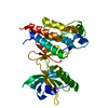
| ||||||||
|---|---|---|---|---|---|---|---|---|---|
| 1 |
| ||||||||
| Unit cell |
|
-Components
| #1: Protein | Mass: 36274.738 Da / Num. of mol.: 1 / Fragment: KINASE DOMAIN / Mutation: E990V Source method: isolated from a genetically manipulated source Details: PROTEIN WAS AUTOPHOSPHORYLATED AT RESIDUE 1059 PRIOR TO CRYSTALLIZATION. THIS PROTEIN CONSTRUCT CONTAINS THE KINASE DOMAIN WITH 50 RESIDUES (RESIDUES 940- 989) OF THE KINASE INSERT DOMAIN ...Details: PROTEIN WAS AUTOPHOSPHORYLATED AT RESIDUE 1059 PRIOR TO CRYSTALLIZATION. THIS PROTEIN CONSTRUCT CONTAINS THE KINASE DOMAIN WITH 50 RESIDUES (RESIDUES 940- 989) OF THE KINASE INSERT DOMAIN DELETED. THE PROTEIN ALSO CONTAINS A GLUTAMIC ACID TO VALINE MUTATION AT RESIDUE 990. Source: (gene. exp.) Homo sapiens (human) / Tissue: AORTA / Production host:   Spodoptera frugiperda (fall armyworm) / References: UniProt: P35968, EC: 2.7.1.112 Spodoptera frugiperda (fall armyworm) / References: UniProt: P35968, EC: 2.7.1.112 |
|---|---|
| #2: Water | ChemComp-HOH /  Mass: 18.015 Da / Num. of mol.: 141 / Source method: isolated from a natural source / Formula: H2O Mass: 18.015 Da / Num. of mol.: 141 / Source method: isolated from a natural source / Formula: H2O |
| Has protein modification | N |
-Experimental details
-Experiment
| Experiment | Method: X-RAY DIFFRACTION / Number of used crystals: 1 |
|---|
- Sample preparation
| Crystal | Density Matthews: 2.41 Å3/Da / Density % sol: 49.01 % | ||||||||||||||||||||||||||||||||||||
|---|---|---|---|---|---|---|---|---|---|---|---|---|---|---|---|---|---|---|---|---|---|---|---|---|---|---|---|---|---|---|---|---|---|---|---|---|---|
| Crystal grow | pH: 7.2 / Details: pH 7.2 | ||||||||||||||||||||||||||||||||||||
| Crystal grow | *PLUS Temperature: 4 ℃ / Method: vapor diffusion, hanging dropDetails: drop consists of equal volume of protein and mother-liquor | ||||||||||||||||||||||||||||||||||||
| Components of the solutions | *PLUS
|
-Data collection
| Diffraction | Mean temperature: 100 K |
|---|---|
| Diffraction source | Source: ROTATING ANODE / Type: RIGAKU RU200 / Wavelength: 1.5418 |
| Detector | Type: MARRESEARCH / Detector: IMAGE PLATE |
| Radiation | Protocol: SINGLE WAVELENGTH / Monochromatic (M) / Laue (L): M / Scattering type: x-ray |
| Radiation wavelength | Wavelength: 1.5418 Å / Relative weight: 1 |
| Reflection | Resolution: 2.2→15 Å / Num. obs: 17234 / % possible obs: 93 % / Redundancy: 3.9 % / Rsym value: 0.052 / Net I/σ(I): 23.6 |
| Reflection shell | Resolution: 2.2→2.28 Å / Mean I/σ(I) obs: 3.4 / Rsym value: 0.196 / % possible all: 81.5 |
| Reflection | *PLUS Rmerge(I) obs: 0.052 |
| Reflection shell | *PLUS % possible obs: 81.5 % / Rmerge(I) obs: 0.196 |
- Processing
| Software |
| ||||||||||||||||||||||||||||||||||||||||||||||||||||||||||||
|---|---|---|---|---|---|---|---|---|---|---|---|---|---|---|---|---|---|---|---|---|---|---|---|---|---|---|---|---|---|---|---|---|---|---|---|---|---|---|---|---|---|---|---|---|---|---|---|---|---|---|---|---|---|---|---|---|---|---|---|---|---|
| Refinement | Method to determine structure: MOLECULAR REPLACEMENT Starting model: PDB ENTRY 1FGK Resolution: 2.4→8 Å / Data cutoff high absF: 10000 / Data cutoff low absF: 1.1 / σ(F): 2
| ||||||||||||||||||||||||||||||||||||||||||||||||||||||||||||
| Displacement parameters | Biso mean: 31.8 Å2 | ||||||||||||||||||||||||||||||||||||||||||||||||||||||||||||
| Refinement step | Cycle: LAST / Resolution: 2.4→8 Å
| ||||||||||||||||||||||||||||||||||||||||||||||||||||||||||||
| Refine LS restraints |
| ||||||||||||||||||||||||||||||||||||||||||||||||||||||||||||
| Xplor file | Serial no: 1 / Param file: PARHCSDX.PRO / Topol file: TOPHCSDX.PRO | ||||||||||||||||||||||||||||||||||||||||||||||||||||||||||||
| Software | *PLUS Name: X-PLOR / Version: 3.1 / Classification: refinement | ||||||||||||||||||||||||||||||||||||||||||||||||||||||||||||
| Refine LS restraints | *PLUS
|
