Oxidoreductases; Acting on the CH-CH group of donors; With unknown physiological acceptors / 3-methylbutanoyl-CoA dehydrogenase activity / acyl-CoA dehydrogenase activity / sequence-specific DNA binding / negative regulation of DNA-templated transcription / DNA damage response / DNA binding / identical protein binding / cytoplasm Similarity search - Function
Butyryl-CoA Dehydrogenase, subunit A; domain 2 - #20 / Single alpha-helices involved in coiled-coils or other helix-helix interfaces - #600 / Putative acyl-CoA dehydrogenase AidB / Adaptive response protein AidB, N-terminal / : / Adaptive response protein AidB N-terminal domain / Acyl-CoA dehydrogenases signature 1. / Acyl-CoA dehydrogenases signature 2. / Acyl-CoA dehydrogenase, conserved site / Butyryl-CoA Dehydrogenase, subunit A; domain 2 ...Butyryl-CoA Dehydrogenase, subunit A; domain 2 - #20 / Single alpha-helices involved in coiled-coils or other helix-helix interfaces - #600 / Putative acyl-CoA dehydrogenase AidB / Adaptive response protein AidB, N-terminal / : / Adaptive response protein AidB N-terminal domain / Acyl-CoA dehydrogenases signature 1. / Acyl-CoA dehydrogenases signature 2. / Acyl-CoA dehydrogenase, conserved site / Butyryl-CoA Dehydrogenase, subunit A; domain 2 / Acyl-CoA dehydrogenase/oxidase C-terminal / Acyl-CoA dehydrogenase, C-terminal domain / Acyl-CoA oxidase/dehydrogenase, middle domain / Acyl-CoA dehydrogenase, middle domain / Acyl-CoA dehydrogenase/oxidase, N-terminal and middle domain superfamily / Acyl-CoA dehydrogenase-like, C-terminal / Single alpha-helices involved in coiled-coils or other helix-helix interfaces / Helix non-globular / Special / Beta Barrel / Mainly Beta Similarity search - Domain/homology
Mass: 18.015 Da / Num. of mol.: 439 / Source method: isolated from a natural source / Formula: H2O
Has protein modification
Y
-
Experimental details
-
Experiment
Experiment
Method: X-RAY DIFFRACTION / Number of used crystals: 1
-
Sample preparation
Crystal
Density Matthews: 2.46 Å3/Da / Density % sol: 49.97 %
Crystal grow
Temperature: 298 K / Method: vapor diffusion, sitting drop / pH: 8 Details: 100 mM Tris pH 8.0, 50 mM CaCl2, and 4% PEG 2000, VAPOR DIFFUSION, SITTING DROP, temperature 298.0K
In the structure databanks used in Yorodumi, some data are registered as the other names, "COVID-19 virus" and "2019-nCoV". Here are the details of the virus and the list of structure data.
Jan 31, 2019. EMDB accession codes are about to change! (news from PDBe EMDB page)
EMDB accession codes are about to change! (news from PDBe EMDB page)
The allocation of 4 digits for EMDB accession codes will soon come to an end. Whilst these codes will remain in use, new EMDB accession codes will include an additional digit and will expand incrementally as the available range of codes is exhausted. The current 4-digit format prefixed with “EMD-” (i.e. EMD-XXXX) will advance to a 5-digit format (i.e. EMD-XXXXX), and so on. It is currently estimated that the 4-digit codes will be depleted around Spring 2019, at which point the 5-digit format will come into force.
The EM Navigator/Yorodumi systems omit the EMD- prefix.
Related info.:Q: What is EMD? / ID/Accession-code notation in Yorodumi/EM Navigator
Yorodumi is a browser for structure data from EMDB, PDB, SASBDB, etc.
This page is also the successor to EM Navigator detail page, and also detail information page/front-end page for Omokage search.
The word "yorodu" (or yorozu) is an old Japanese word meaning "ten thousand". "mi" (miru) is to see.
Related info.:EMDB / PDB / SASBDB / Comparison of 3 databanks / Yorodumi Search / Aug 31, 2016. New EM Navigator & Yorodumi / Yorodumi Papers / Jmol/JSmol / Function and homology information / Changes in new EM Navigator and Yorodumi
 Movie
Movie Controller


 Open data
 Basic information
 Components
 Keywords
 Function and homology information
 X-RAY DIFFRACTION /
  Authors
 Citation
 Structure visualization
 Downloads & links
 Other downloads
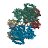


 PDBj
PDBj Assembly


 Mass: 40.078 Da / Num. of mol.: 1 / Source method: obtained synthetically / Formula: Ca
 Mass: 785.550 Da / Num. of mol.: 1 / Source method: obtained synthetically / Formula: C27H33N9O15P2 / Comment: FAD*YM
 Mass: 18.015 Da / Num. of mol.: 439 / Source method: isolated from a natural source / Formula: H2O
 Sample preparation
 / Beamline: 22-ID / Wavelength: 1.02891 Å
 Processing