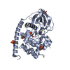
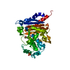
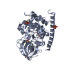
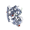
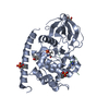
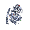
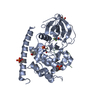
登録情報 データベース : PDB / ID : 3dd2タイトル Crystal structure of an RNA aptamer bound to human thrombin RNA (26-MER) Thrombin heavy chain Thrombin light chain キーワード / / / / / / / / / 機能・相同性 分子機能 ドメイン・相同性 構成要素
/ / / / / / / / / / / / / / / / / / / / / / / / / / / / / / / / / / / / / / / / / / / / / / / / / / / / / / / / / / / / / / / / / / / / / / / / / / / / / / / / / / / / / / / / / / / / / / / / / / / / / / / / / / / / / / / / / / / / / / / 生物種 Homo sapiens (ヒト)手法 / 解像度 : 1.9 Å データ登録者 Long, S.B. / Sullenger, B.A. ジャーナル : Rna / 年 : 2008タイトル : Crystal structure of an RNA aptamer bound to thrombin.著者 : Long, S.B. / Long, M.B. / White, R.R. / Sullenger, B.A. 履歴 登録 2008年6月4日 登録サイト / 処理サイト 改定 1.0 2008年11月11日 Provider / タイプ 改定 1.1 2011年7月13日 Group Atomic model / Database references ... Atomic model / Database references / Derived calculations / Non-polymer description / Structure summary / Version format compliance 改定 1.2 2013年2月27日 Group 改定 1.3 2017年10月25日 Group / カテゴリ 改定 1.4 2023年8月30日 Group Data collection / Database references ... Data collection / Database references / Derived calculations / Refinement description カテゴリ chem_comp_atom / chem_comp_bond ... chem_comp_atom / chem_comp_bond / database_2 / pdbx_initial_refinement_model / pdbx_struct_conn_angle / struct_conn / struct_site Item _database_2.pdbx_DOI / _database_2.pdbx_database_accession ... _database_2.pdbx_DOI / _database_2.pdbx_database_accession / _pdbx_struct_conn_angle.ptnr1_auth_asym_id / _pdbx_struct_conn_angle.ptnr1_auth_comp_id / _pdbx_struct_conn_angle.ptnr1_auth_seq_id / _pdbx_struct_conn_angle.ptnr1_label_asym_id / _pdbx_struct_conn_angle.ptnr1_label_atom_id / _pdbx_struct_conn_angle.ptnr1_label_comp_id / _pdbx_struct_conn_angle.ptnr1_label_seq_id / _pdbx_struct_conn_angle.ptnr2_auth_asym_id / _pdbx_struct_conn_angle.ptnr2_auth_seq_id / _pdbx_struct_conn_angle.ptnr2_label_asym_id / _pdbx_struct_conn_angle.ptnr3_auth_asym_id / _pdbx_struct_conn_angle.ptnr3_auth_comp_id / _pdbx_struct_conn_angle.ptnr3_auth_seq_id / _pdbx_struct_conn_angle.ptnr3_label_asym_id / _pdbx_struct_conn_angle.ptnr3_label_atom_id / _pdbx_struct_conn_angle.ptnr3_label_comp_id / _pdbx_struct_conn_angle.ptnr3_label_seq_id / _pdbx_struct_conn_angle.value / _struct_conn.conn_type_id / _struct_conn.id / _struct_conn.pdbx_dist_value / _struct_conn.pdbx_leaving_atom_flag / _struct_conn.ptnr1_auth_asym_id / _struct_conn.ptnr1_auth_comp_id / _struct_conn.ptnr1_auth_seq_id / _struct_conn.ptnr1_label_asym_id / _struct_conn.ptnr1_label_atom_id / _struct_conn.ptnr1_label_comp_id / _struct_conn.ptnr1_label_seq_id / _struct_conn.ptnr2_auth_asym_id / _struct_conn.ptnr2_auth_comp_id / _struct_conn.ptnr2_auth_seq_id / _struct_conn.ptnr2_label_asym_id / _struct_conn.ptnr2_label_atom_id / _struct_conn.ptnr2_label_comp_id / _struct_conn.ptnr2_label_seq_id / _struct_site.pdbx_auth_asym_id / _struct_site.pdbx_auth_comp_id / _struct_site.pdbx_auth_seq_id 改定 1.5 2024年11月20日 Group カテゴリ / pdbx_modification_featureItem
すべて表示 表示を減らす
 ムービー
ムービー コントローラー


 データを開く
 基本情報
基本情報 要素
要素 キーワード
キーワード 機能・相同性情報
 Homo sapiens (ヒト)
 X線回折 / 解像度: 1.9 Å
  データ登録者
 引用
引用 構造の表示
構造の表示 ダウンロードとリンク
 その他のダウンロード
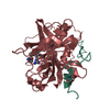


 PDBj
PDBj


 集合体
集合体


 分子量: 106.120 Da / 分子数: 3 / 由来タイプ: 合成 / 式: C4H10O3
 タイプ: peptide-like, Peptide-like / クラス: 阻害剤 / 分子量: 453.986 Da / 分子数: 1 / 由来タイプ: 合成 / 式: C21H34ClN6O3 / 参照: D-Phe-Pro-Arg-CH2Cl
 分子量: 24.305 Da / 分子数: 2 / 由来タイプ: 合成 / 式: Mg
 分子量: 282.331 Da / 分子数: 1 / 由来タイプ: 合成 / 式: C12H26O7 / コメント: 沈殿剤*YM
 分子量: 60.052 Da / 分子数: 6 / 由来タイプ: 合成 / 式: C2H4O2
 試料調製
試料調製 解析
解析