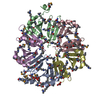
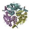
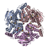
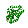
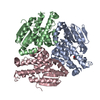
A: Protein of Unknown Function (DUF1696) with Pleckstrin-homology Domains B: Protein of Unknown Function (DUF1696) with Pleckstrin-homology Domains C: Protein of Unknown Function (DUF1696) with Pleckstrin-homology Domains D: Protein of Unknown Function (DUF1696) with Pleckstrin-homology Domains E: Protein of Unknown Function (DUF1696) with Pleckstrin-homology Domains hetero molecules
Component-ID: 1 / Ens-ID: 1 / End auth comp-ID: LEU / End label comp-ID: LEU / Refine code: 4
Dom-ID
Beg auth comp-ID
Beg label comp-ID
Auth asym-ID
Label asym-ID
Auth seq-ID
Label seq-ID
1
GLY
GLY
A
A
9 - 124
2 - 117
2
ALA
ALA
B
B
12 - 124
5 - 117
3
ASN
ASN
C
C
10 - 124
3 - 117
4
ALA
ALA
D
D
12 - 124
5 - 117
5
GLU
GLU
E
E
13 - 124
6 - 117
Details
AUTHORS STATE THAT SIZE EXCLUSION CHROMATOGRAPHY WITH STATIC LIGHT SCATTERING SUPPORTS THE ASSIGNMENT OF A PENTAMER AS A SIGNIFICANT OLIGOMERIZATION STATE IN SOLUTION.
Mass: 18.015 Da / Num. of mol.: 322 / Source method: isolated from a natural source / Formula: H2O
Has protein modification
Y
Sequence details
THE CONSTRUCT WAS EXPRESSED WITH A PURIFICATION TAG MGSDKIHHHHHHENLYFQG. THE TAG WAS REMOVED WITH ...THE CONSTRUCT WAS EXPRESSED WITH A PURIFICATION TAG MGSDKIHHHHHHENLYFQG. THE TAG WAS REMOVED WITH TEV PROTEASE LEAVING ONLY A GLYCINE (0) FOLLOWED BY THE TARGET SEQUENCE. THE CLONED CONSTRUCT CONTAINS RESIDUES 9-124 OF THE FULL LENGTH PROTEIN.
-
Experimental details
-
Experiment
Experiment
Method: X-RAY DIFFRACTION / Number of used crystals: 1
-
Sample preparation
Crystal
Density Matthews: 2.45 Å3/Da / Density % sol: 49.7 %
Crystal grow
Temperature: 277 K / Method: vapor diffusion, sitting drop / pH: 6.83 Details: 37.0% 2-methyl-2,4-pentanediol, 0.15M sodium chloride, 0.1M HEPES pH 6.83, NANODROP, VAPOR DIFFUSION, SITTING DROP, temperature 277K
Monochromator: Single crystal Si(111) bent monochromator (horizontal focusing) Protocol: MAD / Monochromatic (M) / Laue (L): M / Scattering type: x-ray
Radiation wavelength
ID
Wavelength (Å)
Relative weight
1
0.97925
1
2
0.91837
1
3
0.97871
1
Reflection
Resolution: 2→29.828 Å / Num. obs: 43831 / % possible obs: 97.9 % / Observed criterion σ(I): -3 / Redundancy: 4.05 % / Biso Wilson estimate: 31.07 Å2 / Rmerge(I) obs: 0.056 / Net I/σ(I): 11.19
Reflection shell
Resolution (Å)
Rmerge(I) obs
Mean I/σ(I) obs
Num. measured obs
Num. unique obs
Diffraction-ID
% possible all
2-2.07
0.638
1.5
16853
7885
1
95.9
2.07-2.15
0.469
2
17146
7960
1
97.2
2.15-2.25
0.346
2.8
18168
8425
1
98
2.25-2.37
0.268
3.5
17920
8315
1
97.9
2.37-2.52
0.213
4.3
18021
8341
1
98.2
2.52-2.71
0.146
6.2
17473
8085
1
98.3
2.71-2.99
0.092
9.6
18401
8508
1
98.4
2.99-3.42
0.048
16.5
17721
8189
1
98.5
3.42-4.3
0.026
27.7
17874
8232
1
98.5
4.3-29.83
0.019
37.1
18127
8307
1
97.9
-
Phasing
Phasing
Method: MAD
-
Processing
Software
Name
Version
Classification
NB
REFMAC
5.2.0019
refinement
PHENIX
refinement
SHELX
phasing
MolProbity
3beta29
modelbuilding
XSCALE
datascaling
PDB_EXTRACT
3.004
dataextraction
XDS
datareduction
SHELXD
phasing
autoSHARP
phasing
Refinement
Method to determine structure: MAD / Resolution: 2→29.828 Å / Cor.coef. Fo:Fc: 0.96 / Cor.coef. Fo:Fc free: 0.939 / SU B: 7.891 / SU ML: 0.113 / TLS residual ADP flag: LIKELY RESIDUAL / Cross valid method: THROUGHOUT / σ(F): 0 / ESU R: 0.176 / ESU R Free: 0.159 Stereochemistry target values: MAXIMUM LIKELIHOOD WITH PHASES Details: 1. HYDROGENS HAVE BEEN ADDED IN THE RIDING POSITIONS. 2. A MET-INHIBITION PROTOCOL WAS USED FOR SELENOMETHIONINE INCORPORATION DURING PROTEIN EXPRESSION. THE OCCUPANCY OF THE SE ATOMS IN THE ...Details: 1. HYDROGENS HAVE BEEN ADDED IN THE RIDING POSITIONS. 2. A MET-INHIBITION PROTOCOL WAS USED FOR SELENOMETHIONINE INCORPORATION DURING PROTEIN EXPRESSION. THE OCCUPANCY OF THE SE ATOMS IN THE MSE RESIDUES WAS REDUCED TO 0.75 FOR THE REDUCED SCATTERING POWER DUE TO PARTIAL S-MET INCORPORATION. 3. ATOM RECORDS CONTAIN RESIDUAL B FACTORS ONLY. 4. MPD AND CL ARE PRESENT IN CRYSTALLIZATION CONDITION.
Rfactor
Num. reflection
% reflection
Selection details
Rfree
0.227
2200
5 %
RANDOM
Rwork
0.184
-
-
-
obs
0.186
43782
98.93 %
-
Solvent computation
Ion probe radii: 0.8 Å / Shrinkage radii: 0.8 Å / VDW probe radii: 1.2 Å / Solvent model: MASK
In the structure databanks used in Yorodumi, some data are registered as the other names, "COVID-19 virus" and "2019-nCoV". Here are the details of the virus and the list of structure data.
Jan 31, 2019. EMDB accession codes are about to change! (news from PDBe EMDB page)
EMDB accession codes are about to change! (news from PDBe EMDB page)
The allocation of 4 digits for EMDB accession codes will soon come to an end. Whilst these codes will remain in use, new EMDB accession codes will include an additional digit and will expand incrementally as the available range of codes is exhausted. The current 4-digit format prefixed with “EMD-” (i.e. EMD-XXXX) will advance to a 5-digit format (i.e. EMD-XXXXX), and so on. It is currently estimated that the 4-digit codes will be depleted around Spring 2019, at which point the 5-digit format will come into force.
The EM Navigator/Yorodumi systems omit the EMD- prefix.
Related info.:Q: What is EMD? / ID/Accession-code notation in Yorodumi/EM Navigator
Yorodumi is a browser for structure data from EMDB, PDB, SASBDB, etc.
This page is also the successor to EM Navigator detail page, and also detail information page/front-end page for Omokage search.
The word "yorodu" (or yorozu) is an old Japanese word meaning "ten thousand". "mi" (miru) is to see.
Related info.:EMDB / PDB / SASBDB / Comparison of 3 databanks / Yorodumi Search / Aug 31, 2016. New EM Navigator & Yorodumi / Yorodumi Papers / Jmol/JSmol / Function and homology information / Changes in new EM Navigator and Yorodumi
 Movie
Movie Controller

 Yorodumi
 Open data
 Basic information
 Components
 Keywords
 Function and homology information
 Shewanella loihica PV-4 (bacteria)
 X-RAY DIFFRACTION /
  Authors
 Citation
 Structure visualization
 Downloads & links
 Other downloads
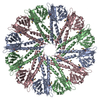


 PDBj
PDBj Assembly

 Mass: 118.174 Da / Num. of mol.: 7 / Source method: obtained synthetically / Formula: C6H14O2 / Comment: precipitant*YM
 Mass: 35.453 Da / Num. of mol.: 1 / Source method: obtained synthetically / Formula: Cl
 Mass: 18.015 Da / Num. of mol.: 322 / Source method: isolated from a natural source / Formula: H2O
 Sample preparation
 / Beamline: BL11-1 / Wavelength: 0.97925,0.91837,0.97871
 Processing