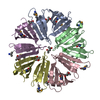
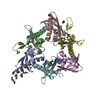
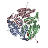
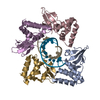
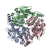
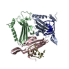
Crystal structure of pleckstrin homology domain (YP_926556.1) from SHEWANELLA AMAZONENSIS SB2B at 1.99 A resolution
Components
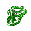
pleckstrin homology domain
Keywords
structural genomics / unknown function / YP_926556.1 / pleckstrin homology domain / Joint Center for Structural Genomics / JCSG / Protein Structure Initiative / PSI-2 / Protein of unknown function (DUF1696)
Mass: 18.015 Da / Num. of mol.: 242 / Source method: isolated from a natural source / Formula: H2O
Sequence details
1. THE CONSTRUCT WAS EXPRESSED WITH A PURIFICATION TAG MGSDKIHHHHHHENLYFQG. THE TAG WAS REMOVED ...1. THE CONSTRUCT WAS EXPRESSED WITH A PURIFICATION TAG MGSDKIHHHHHHENLYFQG. THE TAG WAS REMOVED WITH TEV PROTEASE LEAVING ONLY A GLYCINE (0) FOLLOWED BY THE TARGET SEQUENCE. 2. THE PROTEIN WAS REDUCTIVELY METHYLATED PRIOR TO CRYSTALLIZATION.
-
Experimental details
-
Experiment
Experiment
Method: X-RAY DIFFRACTION / Number of used crystals: 2
-
Sample preparation
Crystal
Density Matthews: 2.07 Å3/Da / Density % sol: 40.61 % Description: THE STRUCTURE WAS SOLVED BY MAD METHOD USING FOUR DATASETS COLLECTED FROM TWO CRYSTALS. THE SHARP OUTPUT PHASES WERE USED AS RESTRAINTS DURING REFINEMENT.
Crystal grow
Temperature: 293 K / Method: vapor diffusion, sitting drop / pH: 7.5 Details: 5.0000% polyethylene glycol 3000, 22.0000% polyethylene glycol 400, 10.0000% Glycerol, 0.1M HEPES pH 7.5, VAPOR DIFFUSION,SITTING DROP,NANODROP, temperature 293K, VAPOR DIFFUSION, SITTING DROP
-
Data collection
Diffraction
ID
Mean temperature (K)
Crystal-ID
1
100
1
2
100
1
Diffraction source
Source
Site
Beamline
ID
Wavelength (Å)
SYNCHROTRON
SSRL
BL9-2
1
0.9792
SYNCHROTRON
SSRL
BL9-2
2
0.97920,0.97934,0.91837
Detector
Type
ID
Detector
Date
Details (eV)
MARMOSAIC 325 mm CCD
1
CCD
May 14, 2009
Flatcollimatingmirror, toroidfocusingmirror
MARMOSAIC 325 mm CCD
2
CCD
May 14, 2009
Flatcollimatingmirror, toroidfocusingmirror
Radiation
ID
Monochromator
Protocol
Monochromatic (M) / Laue (L)
Scattering type
Wavelength-ID
1
Doublecrystalmonochromator
MAD
M
x-ray
1
2
Doublecrystalmonochromator
MAD
M
x-ray
1
Radiation wavelength
ID
Wavelength (Å)
Relative weight
1
0.9792
1
2
0.97934
1
3
0.91837
1
Reflection
Resolution: 1.99→47.351 Å / Num. obs: 41702 / % possible obs: 98.4 % / Observed criterion σ(I): -3 / Redundancy: 3.61 % / Biso Wilson estimate: 22.653 Å2 / Rmerge(I) obs: 0.087 / Net I/σ(I): 10.18
Reflection shell
Resolution (Å)
Rmerge(I) obs
Mean I/σ(I) obs
Num. measured obs
Num. unique obs
Diffraction-ID
% possible all
1.99-2.06
0.531
2.3
14883
4075
1,2
99.1
2.06-2.14
0.408
3
14594
3949
1,2
98.4
2.14-2.24
0.314
3.8
15552
4233
1,2
98.1
2.24-2.36
0.248
4.8
15401
4209
1,2
99.5
2.36-2.51
0.204
5.8
15358
4180
1,2
99
2.51-2.7
0.155
7.6
14852
4067
1,2
98.9
2.7-2.97
0.109
10.3
15139
4180
1,2
99
2.97-3.4
0.071
15
15083
4194
1,2
98.5
3.4-4.27
0.043
22.2
14856
4184
1,2
97.9
4.27-47.351
0.04
25.6
15011
4423
1,2
96
-
Phasing
Phasing
Method: MAD
-
Processing
Software
Name
Version
Classification
NB
REFMAC
5.5.0092
refinement
PHENIX
refinement
SHELX
phasing
MolProbity
3beta29
modelbuilding
XSCALE
datascaling
PDB_EXTRACT
3.006
dataextraction
XDS
datareduction
SHELXD
phasing
autoSHARP
phasing
Refinement
Method to determine structure: MAD / Resolution: 1.99→47.351 Å / Cor.coef. Fo:Fc: 0.948 / Cor.coef. Fo:Fc free: 0.92 / Occupancy max: 1 / Occupancy min: 0.23 / SU B: 8.591 / SU ML: 0.11 / TLS residual ADP flag: LIKELY RESIDUAL / Cross valid method: THROUGHOUT / σ(F): 0 / ESU R: 0.193 / ESU R Free: 0.168 Stereochemistry target values: MAXIMUM LIKELIHOOD WITH PHASES Details: 1. HYDROGENS HAVE BEEN ADDED IN THE RIDING POSITIONS. 2. A MET-INHIBITION PROTOCOL WAS USED FOR SELENOMETHIONINE INCORPORATION DURING PROTEIN EXPRESSION. THE OCCUPANCY OF THE SE ATOMS IN THE ...Details: 1. HYDROGENS HAVE BEEN ADDED IN THE RIDING POSITIONS. 2. A MET-INHIBITION PROTOCOL WAS USED FOR SELENOMETHIONINE INCORPORATION DURING PROTEIN EXPRESSION. THE OCCUPANCY OF THE SE ATOMS IN THE MSE RESIDUES WAS REDUCED TO 0.75 FOR THE REDUCED SCATTERING POWER DUE TO PARTIAL S-MET INCORPORATION. 3. ATOM RECORDS CONTAIN RESIDUAL B FACTORS ONLY. 4. GLYCEROL (GOL) AND POLY ETHYLENE GLYCOL FRAGMENTS (PEG,PGE) MODELED ARE PRESENT IN CRYSTALLIZATION/CRYO CONDITIONS.
Rfactor
Num. reflection
% reflection
Selection details
Rfree
0.233
2115
5.1 %
RANDOM
Rwork
0.189
-
-
-
obs
0.191
41641
98.47 %
-
Solvent computation
Ion probe radii: 0.8 Å / Shrinkage radii: 0.8 Å / VDW probe radii: 1.2 Å / Solvent model: MASK
In the structure databanks used in Yorodumi, some data are registered as the other names, "COVID-19 virus" and "2019-nCoV". Here are the details of the virus and the list of structure data.
Jan 31, 2019. EMDB accession codes are about to change! (news from PDBe EMDB page)
EMDB accession codes are about to change! (news from PDBe EMDB page)
The allocation of 4 digits for EMDB accession codes will soon come to an end. Whilst these codes will remain in use, new EMDB accession codes will include an additional digit and will expand incrementally as the available range of codes is exhausted. The current 4-digit format prefixed with “EMD-” (i.e. EMD-XXXX) will advance to a 5-digit format (i.e. EMD-XXXXX), and so on. It is currently estimated that the 4-digit codes will be depleted around Spring 2019, at which point the 5-digit format will come into force.
The EM Navigator/Yorodumi systems omit the EMD- prefix.
Related info.:Q: What is EMD? / ID/Accession-code notation in Yorodumi/EM Navigator
Yorodumi is a browser for structure data from EMDB, PDB, SASBDB, etc.
This page is also the successor to EM Navigator detail page, and also detail information page/front-end page for Omokage search.
The word "yorodu" (or yorozu) is an old Japanese word meaning "ten thousand". "mi" (miru) is to see.
Related info.:EMDB / PDB / SASBDB / Comparison of 3 databanks / Yorodumi Search / Aug 31, 2016. New EM Navigator & Yorodumi / Yorodumi Papers / Jmol/JSmol / Function and homology information / Changes in new EM Navigator and Yorodumi
 Movie
Movie Controller

 Yorodumi
 Open data
 Basic information
 Components
 Keywords
 Function and homology information
 Shewanella amazonensis SB2B (bacteria)
 X-RAY DIFFRACTION /
  Authors
 Citation
 Structure visualization
 Downloads & links
 Other downloads
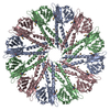


 PDBj
PDBj Assembly
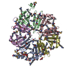

 Mass: 92.094 Da / Num. of mol.: 6 / Source method: obtained synthetically / Formula: C3H8O3
 Mass: 106.120 Da / Num. of mol.: 2 / Source method: obtained synthetically / Formula: C4H10O3
 Mass: 18.015 Da / Num. of mol.: 242 / Source method: isolated from a natural source / Formula: H2O
 Sample preparation
 Processing