 Movie
Movie Controller

[English] 日本語
 Yorodumi
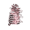
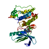
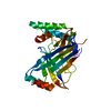
- PDB-3dca: Crystal structure of the RPA0582- protein of unknown function fro... -
+ Open data

- Basic information
| Entry | Database: PDB / ID: 3dca | ||||||
|---|---|---|---|---|---|---|---|
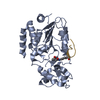
| Title | Crystal structure of the RPA0582- protein of unknown function from Rhodopseudomonas palustris- a structural genomics target | ||||||
 Components Components | RPA0582 | ||||||
 Keywords Keywords | structural genomics / unknown function / Alpha-beta-barrel / SAD / PSI-2 / Protein Structure Initiative / Midwest Center for Structural Genomics / MCSG | ||||||
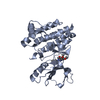
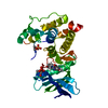
| Function / homology | Domain of unknown function DUF1330 / Domain of unknown function (DUF1330) / Alpha-Beta Plaits - #100 / Dimeric alpha-beta barrel / Alpha-Beta Plaits / 2-Layer Sandwich / Alpha Beta / DUF1330 domain-containing protein Function and homology information Function and homology information | ||||||
| Biological species |  Rhodopseudomonas palustris (phototrophic) Rhodopseudomonas palustris (phototrophic) | ||||||
| Method |  X-RAY DIFFRACTION / SYNCHROTRON / SAD / Resolution: 3.35 Å X-RAY DIFFRACTION / SYNCHROTRON / SAD / Resolution: 3.35 Å | ||||||
 Authors Authors | Sledz, P. / Wang, S. / Chruszcz, M. / Yim, V. / Kudritska, M. / Evdokimova, E. / Turk, D. / Savchenko, A. / Edwards, A. / Joachimiak, A. ...Sledz, P. / Wang, S. / Chruszcz, M. / Yim, V. / Kudritska, M. / Evdokimova, E. / Turk, D. / Savchenko, A. / Edwards, A. / Joachimiak, A. / Minor, W. / Midwest Center for Structural Genomics (MCSG) | ||||||
 Citation Citation | Journal: To be Published Title: Crystal structure of the RPA0582- protein of unknown function from Rhodopseudomonas palustris- a structural genomics target Authors: Sledz, P. / Wang, S. / Chruszcz, M. / Yim, V. / Kudritska, M. / Evdokimova, E. / Turk, D. / Savchenko, A. / Edwards, A. / Minor, W. | ||||||
| History |
|
- Structure visualization
| Structure viewer | Molecule: MolmilJmol/JSmol |
|---|
- Downloads & links
-Download
| PDBx/mmCIF format | 3dca.cif.gz | 107.6 KB | Display | PDBx/mmCIF format |
|---|---|---|---|---|
| PDB format | pdb3dca.ent.gz | 83.4 KB | Display | PDB format |
| PDBx/mmJSON format | 3dca.json.gz | Tree view | PDBx/mmJSON format | |
| Others |  Other downloads Other downloads |
-Validation report
| Arichive directory | https://data.pdbj.org/pub/pdb/validation_reports/dc/3dcaftp://data.pdbj.org/pub/pdb/validation_reports/dc/3dca | HTTPS FTP |
|---|
-Related structure data
| Similar structure data | |
|---|---|
| Other databases |

-Links
 PDBj
PDBj- Assembly
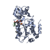
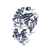
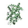
| Deposited unit | 
| ||||||||||||||||||||||||||||||
|---|---|---|---|---|---|---|---|---|---|---|---|---|---|---|---|---|---|---|---|---|---|---|---|---|---|---|---|---|---|---|---|
| 1 | 
| ||||||||||||||||||||||||||||||
| 2 | 
| ||||||||||||||||||||||||||||||
| Unit cell |
| ||||||||||||||||||||||||||||||
| Components on special symmetry positions |
| ||||||||||||||||||||||||||||||
| Noncrystallographic symmetry (NCS) | NCS domain:
NCS domain segments: Component-ID: 1 / Ens-ID: 1 / Beg auth comp-ID: THR / Beg label comp-ID: THR / End auth comp-ID: PRO / End label comp-ID: PRO / Refine code: 1 / Auth seq-ID: 2 - 133 / Label seq-ID: 2 - 133
|
-Components
| #1: Protein | Mass: 16269.880 Da / Num. of mol.: 4 Source method: isolated from a genetically manipulated source Source: (gene. exp.) Rhodopseudomonas palustris (phototrophic)Gene: RPA0582 / Plasmid: P15TV LIC / Production host: #2: Chemical | ChemComp-SO4 /   Mass: 96.063 Da / Num. of mol.: 6 / Source method: obtained synthetically / Formula: SO4 Mass: 96.063 Da / Num. of mol.: 6 / Source method: obtained synthetically / Formula: SO4Has protein modification | Y | |
|---|
-Experimental details
-Experiment
| Experiment | Method: X-RAY DIFFRACTION / Number of used crystals: 1 |
|---|
- Sample preparation
| Crystal | Density Matthews: 6.58 Å3/Da / Density % sol: 81.3 % |
|---|---|
| Crystal grow | Temperature: 293 K / Method: vapor diffusion, hanging drop / pH: 6.5 Details: 0.2M AMMONIUM SULPHATE, 0.1M NA CACODILATE PH6.5, 30%PEG8K, , VAPOR DIFFUSION, HANGING DROP, temperature 293K |
-Data collection
| Diffraction | Mean temperature: 100 K | |||||||||
|---|---|---|---|---|---|---|---|---|---|---|
| Diffraction source | Source: SYNCHROTRON / Site: APS  / Beamline: 19-ID / Wavelength: 0.97918 / Wavelength: 0.979 Å / Beamline: 19-ID / Wavelength: 0.97918 / Wavelength: 0.979 Å | |||||||||
| Detector | Type: ADSC QUANTUM 315 / Detector: CCD / Date: Feb 10, 2007 / Details: MIRROR | |||||||||
| Radiation | Monochromator: SI-111 CHANNEL / Protocol: SINGLE WAVELENGTH / Monochromatic (M) / Laue (L): M / Scattering type: x-ray | |||||||||
| Radiation wavelength |
| |||||||||
| Reflection | Resolution: 3.35→36.59 Å / Num. all: 22256 / Num. obs: 22256 / % possible obs: 93.6 % / Observed criterion σ(F): 0 / Observed criterion σ(I): -3 / Redundancy: 2 % / Biso Wilson estimate: 98.16 Å2 / Rmerge(I) obs: 0.088 / Rsym value: 0.088 / Net I/σ(I): 13.716 | |||||||||
| Reflection shell | Resolution: 3.35→3.47 Å / Redundancy: 2 % / Rmerge(I) obs: 0.426 / Mean I/σ(I) obs: 1.5 / Num. unique all: 2257 / Rsym value: 0.426 / % possible all: 93.2 |
- Processing
| Software |
| ||||||||||||||||||||||||||||||||||||||||||||||||||||||||||||||||||||||||||||||||||||||||||||||||||||||||||||||||||||||||||||||||||||||||||||||||||||||||||||||||||||||||||
|---|---|---|---|---|---|---|---|---|---|---|---|---|---|---|---|---|---|---|---|---|---|---|---|---|---|---|---|---|---|---|---|---|---|---|---|---|---|---|---|---|---|---|---|---|---|---|---|---|---|---|---|---|---|---|---|---|---|---|---|---|---|---|---|---|---|---|---|---|---|---|---|---|---|---|---|---|---|---|---|---|---|---|---|---|---|---|---|---|---|---|---|---|---|---|---|---|---|---|---|---|---|---|---|---|---|---|---|---|---|---|---|---|---|---|---|---|---|---|---|---|---|---|---|---|---|---|---|---|---|---|---|---|---|---|---|---|---|---|---|---|---|---|---|---|---|---|---|---|---|---|---|---|---|---|---|---|---|---|---|---|---|---|---|---|---|---|---|---|---|---|---|
| Refinement | Method to determine structure: SAD / Resolution: 3.35→36.59 Å / Cor.coef. Fo:Fc: 0.899 / Cor.coef. Fo:Fc free: 0.876 / Cross valid method: THROUGHOUT / σ(F): 0 / σ(I): 0 / ESU R: 0.615 / ESU R Free: 0.437 / Stereochemistry target values: MAXIMUM LIKELIHOOD / Details: HYDROGENS HAVE BEEN ADDED IN THE
| ||||||||||||||||||||||||||||||||||||||||||||||||||||||||||||||||||||||||||||||||||||||||||||||||||||||||||||||||||||||||||||||||||||||||||||||||||||||||||||||||||||||||||
| Solvent computation | Ion probe radii: 0.8 Å / Shrinkage radii: 0.8 Å / VDW probe radii: 1.2 Å / Solvent model: MASK | ||||||||||||||||||||||||||||||||||||||||||||||||||||||||||||||||||||||||||||||||||||||||||||||||||||||||||||||||||||||||||||||||||||||||||||||||||||||||||||||||||||||||||
| Displacement parameters | Biso mean: 30.57 Å2
| ||||||||||||||||||||||||||||||||||||||||||||||||||||||||||||||||||||||||||||||||||||||||||||||||||||||||||||||||||||||||||||||||||||||||||||||||||||||||||||||||||||||||||
| Refinement step | Cycle: LAST / Resolution: 3.35→36.59 Å
| ||||||||||||||||||||||||||||||||||||||||||||||||||||||||||||||||||||||||||||||||||||||||||||||||||||||||||||||||||||||||||||||||||||||||||||||||||||||||||||||||||||||||||
| Refine LS restraints |
| ||||||||||||||||||||||||||||||||||||||||||||||||||||||||||||||||||||||||||||||||||||||||||||||||||||||||||||||||||||||||||||||||||||||||||||||||||||||||||||||||||||||||||
| Refine LS restraints NCS | Dom-ID: 1 / Ens-ID: 1 / Number: 1525 / Refine-ID: X-RAY DIFFRACTION / Type: tight positional / Weight position: 0.05
| ||||||||||||||||||||||||||||||||||||||||||||||||||||||||||||||||||||||||||||||||||||||||||||||||||||||||||||||||||||||||||||||||||||||||||||||||||||||||||||||||||||||||||
| LS refinement shell | Resolution: 3.35→3.44 Å / Total num. of bins used: 20
|
