 Movie
Movie Controller


+ Open data

- Basic information
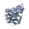
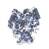
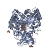
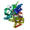
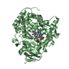
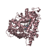
| Entry | Database: PDB / ID: 3dbm | ||||||
|---|---|---|---|---|---|---|---|
| Title | Crystal Structure of Allene oxide synthase | ||||||
 Components Components | Cytochrome P450 74A2 | ||||||
 Keywords Keywords | LYASE / crystal structure HEM HODE AOS / Fatty acid biosynthesis / Heme / Iron / Lipid synthesis / Metal-binding / Oxylipin biosynthesis | ||||||
| Function / homology |  Function and homology information Function and homology informationhydroperoxide dehydratase / allene oxide synthase activity / jasmonic acid biosynthetic process / oxylipin biosynthetic process / sterol metabolic process / chloroplast envelope / chloroplast thylakoid membrane / oxidoreductase activity, acting on paired donors, with incorporation or reduction of molecular oxygen / monooxygenase activity / iron ion binding / heme binding Similarity search - Function | ||||||
| Biological species |  Parthenium argentatum (plant) Parthenium argentatum (plant) | ||||||
| Method |  X-RAY DIFFRACTION / MOLECULAR REPLACEMENT / Resolution: 2.6 Å X-RAY DIFFRACTION / MOLECULAR REPLACEMENT / Resolution: 2.6 Å | ||||||
 Authors Authors | Li, L. / Wang, X. | ||||||
 Citation Citation | Journal: Proc.Natl.Acad.Sci.Usa / Year: 2008 Title: Modes of heme binding and substrate access for cytochrome P450 CYP74A revealed by crystal structures of allene oxide synthase. Authors: Li, L. / Chang, Z. / Pan, Z. / Fu, Z.Q. / Wang, X. | ||||||
| History |
|
- Structure visualization
| Structure viewer | Molecule: MolmilJmol/JSmol |
|---|
- Downloads & links
-Download
| PDBx/mmCIF format | 3dbm.cif.gz | 110.4 KB | Display | PDBx/mmCIF format |
|---|---|---|---|---|
| PDB format | pdb3dbm.ent.gz | 84.1 KB | Display | PDB format |
| PDBx/mmJSON format | 3dbm.json.gz | Tree view | PDBx/mmJSON format | |
| Others |  Other downloads Other downloads |
-Validation report
| Arichive directory | https://data.pdbj.org/pub/pdb/validation_reports/db/3dbmftp://data.pdbj.org/pub/pdb/validation_reports/db/3dbm | HTTPS FTP |
|---|
-Related structure data
| Related structure data |  3damSC  3danC S: Starting model for refinement C: citing same article ( |
|---|---|
| Similar structure data |


-Links
 PDBj
PDBj
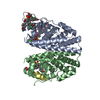
- Assembly
| Deposited unit | 
| ||||||||
|---|---|---|---|---|---|---|---|---|---|
| 1 |
| ||||||||
| Unit cell |
|
-Components
| #1: Protein | Mass: 53532.980 Da / Num. of mol.: 1 Source method: isolated from a genetically manipulated source Source: (gene. exp.) Parthenium argentatum (plant) / Gene: CYP74A2, RPP30 / Production host:  |
|---|---|
| #2: Chemical | ChemComp-HEM /   Mass: 616.487 Da / Num. of mol.: 1 / Source method: obtained synthetically / Formula: C34H32FeN4O4 Mass: 616.487 Da / Num. of mol.: 1 / Source method: obtained synthetically / Formula: C34H32FeN4O4 |
| #3: Chemical | ChemComp-HO2 / (  Mass: 296.445 Da / Num. of mol.: 1 / Source method: obtained synthetically / Formula: C18H32O3 Mass: 296.445 Da / Num. of mol.: 1 / Source method: obtained synthetically / Formula: C18H32O3 |
| #4: Water | ChemComp-HOH /  Mass: 18.015 Da / Num. of mol.: 147 / Source method: isolated from a natural source / Formula: H2O Mass: 18.015 Da / Num. of mol.: 147 / Source method: isolated from a natural source / Formula: H2O |
-Experimental details
-Experiment
| Experiment | Method: X-RAY DIFFRACTION / Number of used crystals: 1 |
|---|
- Sample preparation
| Crystal | Density Matthews: 3.07 Å3/Da / Density % sol: 60 % |
|---|---|
| Crystal grow | Temperature: 277 K / Method: vapor diffusion, hanging drop / pH: 8.5 Details: 0.2M (NH4)H2PO4, 50%MPD, 0.1M Tris pH 8.5, VAPOR DIFFUSION, HANGING DROP, temperature 277K |
-Data collection
| Diffraction | Mean temperature: 100 K |
|---|---|
| Diffraction source | Source: ROTATING ANODE / Type: RIGAKU RUH3R / Wavelength: 1.5418 Å |
| Detector | Type: RIGAKU RAXIS IV++ / Detector: IMAGE PLATE / Date: Apr 20, 2008 |
| Radiation | Protocol: SINGLE WAVELENGTH / Monochromatic (M) / Laue (L): M / Scattering type: x-ray |
| Radiation wavelength | Wavelength: 1.5418 Å / Relative weight: 1 |
| Reflection | Resolution: 2.6→50 Å / Num. all: 20900 / Num. obs: 20482 / % possible obs: 98 % / Redundancy: 98 % / Biso Wilson estimate: 11 Å2 / Rsym value: 0.113 / Net I/σ(I): 10.5 |
| Reflection shell | Resolution: 2.6→2.69 Å / Redundancy: 96 % / Mean I/σ(I) obs: 3.2 / Num. unique all: 1978 / Rsym value: 0.333 / % possible all: 96 |
- Processing
| Software |
| ||||||||||||||||||||
|---|---|---|---|---|---|---|---|---|---|---|---|---|---|---|---|---|---|---|---|---|---|
| Refinement | Method to determine structure: MOLECULAR REPLACEMENT Starting model: 3DAM Resolution: 2.6→49.25 Å / Rfactor Rfree error: 0.006 / Data cutoff high absF: 41885.15 / Data cutoff low absF: 0 / Isotropic thermal model: RESTRAINED / Cross valid method: THROUGHOUT / σ(F): 0 / Stereochemistry target values: Engh & Huber / Details: BULK SOLVENT MODEL USED
| ||||||||||||||||||||
| Solvent computation | Solvent model: FLAT MODEL / Bsol: 30.5769 Å2 / ksol: 0.35 e/Å3 | ||||||||||||||||||||
| Displacement parameters | Biso mean: 27.2 Å2
| ||||||||||||||||||||
| Refine analyze |
| ||||||||||||||||||||
| Refinement step | Cycle: LAST / Resolution: 2.6→49.25 Å
| ||||||||||||||||||||
| Refine LS restraints |
| ||||||||||||||||||||
| LS refinement shell | Resolution: 2.6→2.76 Å / Rfactor Rfree error: 0.019 / Total num. of bins used: 6
| ||||||||||||||||||||
| Xplor file |
|
