 Movie
Movie Controller

[English] 日本語
 Yorodumi
- PDB-3dsi: Crystal Structure of Arabidopsis thaliana Allene Oxide Synthase (... -
+ Open data

- Basic information
| Entry | Database: PDB / ID: 3dsi | ||||||
|---|---|---|---|---|---|---|---|
| Title | Crystal Structure of Arabidopsis thaliana Allene Oxide Synthase (AOS, cytochrome P450 74A, CYP74A) Complexed with 13(S)-HOT at 1.60 A resolution | ||||||
 Components Components | Cytochrome P450 74A, chloroplast | ||||||
 Keywords Keywords | LYASE / P450 / Chloroplast / Fatty acid biosynthesis / Heme / Iron / Lipid synthesis / Metal-binding / Oxylipin biosynthesis / Transit peptide | ||||||
| Function / homology |  Function and homology information Function and homology informationoxylipin metabolic process / hydroperoxide dehydratase / allene oxide synthase activity / plastoglobule / jasmonic acid biosynthetic process / epoxygenase P450 pathway / oxylipin biosynthetic process / response to fungus / chloroplast thylakoid / response to jasmonic acid ...oxylipin metabolic process / hydroperoxide dehydratase / allene oxide synthase activity / plastoglobule / jasmonic acid biosynthetic process / epoxygenase P450 pathway / oxylipin biosynthetic process / response to fungus / chloroplast thylakoid / response to jasmonic acid / thylakoid / hydro-lyase activity / chloroplast envelope / plastid / chloroplast thylakoid membrane / oxidoreductase activity, acting on paired donors, with incorporation or reduction of molecular oxygen / defense response to fungus / chloroplast / monooxygenase activity / defense response / oxygen binding / response to wounding / iron ion binding / heme binding / mitochondrion Similarity search - Function | ||||||
| Biological species |  | ||||||
| Method |  X-RAY DIFFRACTION / SYNCHROTRON / FOURIER SYNTHESIS / Resolution: 1.6 Å X-RAY DIFFRACTION / SYNCHROTRON / FOURIER SYNTHESIS / Resolution: 1.6 Å | ||||||
 Authors Authors | Lee, D.S. / Nioche, P. / Raman, C.S. | ||||||
 Citation Citation | Journal: Nature / Year: 2008 Title: Structural insights into the evolutionary paths of oxylipin biosynthetic enzymes Authors: Lee, D.S. / Nioche, P. / Hamberg, M. / Raman, C.S. | ||||||
| History |
|
- Structure visualization
| Structure viewer | Molecule: MolmilJmol/JSmol |
|---|
- Downloads & links
-Download
| PDBx/mmCIF format | 3dsi.cif.gz | 222.1 KB | Display | PDBx/mmCIF format |
|---|---|---|---|---|
| PDB format | pdb3dsi.ent.gz | 174.2 KB | Display | PDB format |
| PDBx/mmJSON format | 3dsi.json.gz | Tree view | PDBx/mmJSON format | |
| Others |  Other downloads Other downloads |
-Validation report
| Arichive directory | https://data.pdbj.org/pub/pdb/validation_reports/ds/3dsiftp://data.pdbj.org/pub/pdb/validation_reports/ds/3dsi | HTTPS FTP |
|---|
-Related structure data
| Related structure data |  2rchC  2rclC  2rcmC  3cliSC  3dsjC  3dskC C: citing same article ( S: Starting model for refinement |
|---|---|
| Similar structure data |


-Links
 PDBj
PDBj

- Assembly
| Deposited unit | 
| ||||||||
|---|---|---|---|---|---|---|---|---|---|
| 1 | 
| ||||||||
| 2 | 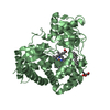
| ||||||||
| Unit cell |
|
-Components
-Protein , 1 types, 2 molecules AB
| #1: Protein | Mass: 55766.590 Da / Num. of mol.: 2 / Fragment: UNP residues 34-518 Source method: isolated from a genetically manipulated source Source: (gene. exp.)  |
|---|
-Non-polymers , 5 types, 815 molecules 


| #2: Chemical |  Mass: 616.487 Da / Num. of mol.: 2 / Source method: obtained synthetically / Formula: C34H32FeN4O4 Mass: 616.487 Da / Num. of mol.: 2 / Source method: obtained synthetically / Formula: C34H32FeN4O4#3: Chemical | ChemComp-T24 / ( |  Mass: 294.429 Da / Num. of mol.: 1 / Source method: obtained synthetically / Formula: C18H30O3 Mass: 294.429 Da / Num. of mol.: 1 / Source method: obtained synthetically / Formula: C18H30O3#4: Chemical |  Mass: 114.229 Da / Num. of mol.: 2 / Source method: obtained synthetically / Formula: C8H18 Mass: 114.229 Da / Num. of mol.: 2 / Source method: obtained synthetically / Formula: C8H18#5: Chemical |  Mass: 92.094 Da / Num. of mol.: 3 / Source method: obtained synthetically / Formula: C3H8O3 Mass: 92.094 Da / Num. of mol.: 3 / Source method: obtained synthetically / Formula: C3H8O3#6: Water | ChemComp-HOH / | Mass: 18.015 Da / Num. of mol.: 807 / Source method: isolated from a natural source / Formula: H2O |
|---|
-Experimental details
-Experiment
| Experiment | Method: X-RAY DIFFRACTION / Number of used crystals: 1 |
|---|
- Sample preparation
| Crystal | Density Matthews: 2.42 Å3/Da / Density % sol: 49.18 % |
|---|---|
| Crystal grow | Temperature: 293 K / Method: vapor diffusion, sitting drop / pH: 7.5 Details: PEG4000, Glycerol, Tris-HCl, pH7.5, VAPOR DIFFUSION, SITTING DROP, temperature 293K |
-Data collection
| Diffraction | Mean temperature: 100 K |
|---|---|
| Diffraction source | Source: SYNCHROTRON / Site: SSRL  / Beamline: BL9-2 / Wavelength: 1.0332 Å / Beamline: BL9-2 / Wavelength: 1.0332 Å |
| Detector | Type: MARMOSAIC 325 mm CCD / Detector: CCD / Date: Aug 4, 2007 |
| Radiation | Monochromator: double miror / Protocol: SINGLE WAVELENGTH / Monochromatic (M) / Laue (L): M / Scattering type: x-ray |
| Radiation wavelength | Wavelength: 1.0332 Å / Relative weight: 1 |
| Reflection | Resolution: 1.6→100 Å / Num. all: 143678 / Num. obs: 143678 / % possible obs: 99.6 % / Observed criterion σ(I): 0 |
| Reflection shell | Resolution: 1.6→1.63 Å / % possible all: 99.1 |
- Processing
| Software |
| ||||||||||||||||||||||||||||||||||||||||||||||||||||||||||||||||||||||||||||||||||||||||||
|---|---|---|---|---|---|---|---|---|---|---|---|---|---|---|---|---|---|---|---|---|---|---|---|---|---|---|---|---|---|---|---|---|---|---|---|---|---|---|---|---|---|---|---|---|---|---|---|---|---|---|---|---|---|---|---|---|---|---|---|---|---|---|---|---|---|---|---|---|---|---|---|---|---|---|---|---|---|---|---|---|---|---|---|---|---|---|---|---|---|---|---|
| Refinement | Method to determine structure: FOURIER SYNTHESIS Starting model: PDB ENTRY 3CLI Resolution: 1.6→88.04 Å / Cor.coef. Fo:Fc: 0.967 / Cor.coef. Fo:Fc free: 0.962 / SU B: 2.384 / SU ML: 0.045 / TLS residual ADP flag: LIKELY RESIDUAL / Cross valid method: THROUGHOUT / σ(F): 0 / ESU R: 0.079 / ESU R Free: 0.076 / Stereochemistry target values: MAXIMUM LIKELIHOOD
| ||||||||||||||||||||||||||||||||||||||||||||||||||||||||||||||||||||||||||||||||||||||||||
| Solvent computation | Ion probe radii: 0.8 Å / Shrinkage radii: 0.8 Å / VDW probe radii: 1.4 Å / Solvent model: MASK | ||||||||||||||||||||||||||||||||||||||||||||||||||||||||||||||||||||||||||||||||||||||||||
| Displacement parameters | Biso mean: 20.979 Å2
| ||||||||||||||||||||||||||||||||||||||||||||||||||||||||||||||||||||||||||||||||||||||||||
| Refinement step | Cycle: LAST / Resolution: 1.6→88.04 Å
| ||||||||||||||||||||||||||||||||||||||||||||||||||||||||||||||||||||||||||||||||||||||||||
| Refine LS restraints |
| ||||||||||||||||||||||||||||||||||||||||||||||||||||||||||||||||||||||||||||||||||||||||||
| LS refinement shell | Resolution: 1.595→1.636 Å / Total num. of bins used: 20
| ||||||||||||||||||||||||||||||||||||||||||||||||||||||||||||||||||||||||||||||||||||||||||
| Refinement TLS params. | Method: refined / Refine-ID: X-RAY DIFFRACTION
| ||||||||||||||||||||||||||||||||||||||||||||||||||||||||||||||||||||||||||||||||||||||||||
| Refinement TLS group |
|
