[English] 日本語
Yorodumi
- PDB-3d9r: Crystal structure of ketosteroid isomerase-like protein (YP_04958... -

+
Open data


ID or keywords:

Loading...

-
Basic information

Entry
Database: PDB / ID: 3d9r
TitleCrystal structure of ketosteroid isomerase-like protein (YP_049581.1) from ERWINIA CAROTOVORA ATROSEPTICA SCRI1043 at 2.40 A resolution
Componentsketosteroid isomerase-like protein
KeywordsISOMERASE / YP_049581.1 / ketosteroid isomerase-like protein / Structural Genomics / Joint Center for Structural Genomics / JCSG / Protein Structure Initiative / PSI-2
Function / homologySnoaL-like domain / SnoaL-like domain / Nuclear Transport Factor 2; Chain: A, - #50 / NTF2-like domain superfamily / Nuclear Transport Factor 2; Chain: A, / Roll / Alpha Beta / Unknown ligand / SnoaL-like domain-containing protein
Function and homology information
Biological speciesPectobacterium atrosepticum (bacteria)
MethodX-RAY DIFFRACTION / SYNCHROTRON / MAD / Resolution: 2.4 Å
AuthorsJoint Center for Structural Genomics (JCSG)
CitationJournal: To be published
Title: Crystal structure of ketosteroid isomerase-like protein (YP_049581.1) from ERWINIA CAROTOVORA ATROSEPTICA SCRI1043 at 2.40 A resolution
Authors: Joint Center for Structural Genomics (JCSG)
History
DepositionMay 27, 2008Deposition site: RCSB / Processing site: RCSB
Revision 1.0Jul 1, 2008Provider: repository / Type: Initial release
Revision 1.1Jul 13, 2011Group: Advisory / Version format compliance
Revision 1.2Oct 25, 2017Group: Author supporting evidence / Refinement description / Category: pdbx_struct_assembly_auth_evidence / software / Item: _software.classification / _software.name
Revision 1.3Jul 24, 2019Group: Data collection / Derived calculations / Refinement description
Category: software / struct_conn
Item: _software.classification / _software.contact_author ..._software.classification / _software.contact_author / _software.contact_author_email / _software.language / _software.location / _software.name / _software.type / _software.version / _struct_conn.pdbx_leaving_atom_flag
Revision 1.4Feb 1, 2023Group: Database references / Derived calculations
Category: database_2 / pdbx_struct_conn_angle ...database_2 / pdbx_struct_conn_angle / struct_conn / struct_ref_seq_dif / struct_site
Item: _database_2.pdbx_DOI / _database_2.pdbx_database_accession ..._database_2.pdbx_DOI / _database_2.pdbx_database_accession / _pdbx_struct_conn_angle.ptnr1_auth_seq_id / _pdbx_struct_conn_angle.ptnr3_auth_seq_id / _pdbx_struct_conn_angle.value / _struct_conn.pdbx_dist_value / _struct_conn.ptnr2_auth_seq_id / _struct_ref_seq_dif.details / _struct_site.pdbx_auth_asym_id / _struct_site.pdbx_auth_comp_id / _struct_site.pdbx_auth_seq_id

-
Structure visualization

Structure viewerMolecule:
MolmilJmol/JSmol

Downloads & links

-
Assembly

Deposited unit
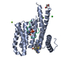
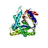
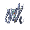
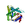
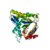
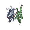
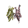
A: ketosteroid isomerase-like protein
B: ketosteroid isomerase-like protein
C: ketosteroid isomerase-like protein
D: ketosteroid isomerase-like protein
hetero molecules


Theoretical massNumber of molelcules
Total (without water)59,10413
Polymers58,7124
Non-polymers3919
Water3,873215
1
A: ketosteroid isomerase-like protein
B: ketosteroid isomerase-like protein
hetero molecules


Theoretical massNumber of molelcules
Total (without water)29,6558
Polymers29,3562
Non-polymers2996
Water362
TypeNameSymmetry operationNumber
identity operation1_555x,y,z1
Buried area3600 Å2
ΔGint-31 kcal/mol
Surface area12260 Å2
MethodPISA
2
C: ketosteroid isomerase-like protein
D: ketosteroid isomerase-like protein
hetero molecules


Theoretical massNumber of molelcules
Total (without water)29,4485
Polymers29,3562
Non-polymers923
Water362
TypeNameSymmetry operationNumber
identity operation1_555x,y,z1
Buried area3320 Å2
ΔGint-20 kcal/mol
Surface area12200 Å2
MethodPISA
Unit cell
Length a, b, c (Å)115.998, 135.416, 139.164
Angle α, β, γ (deg.)90.000, 90.000, 90.000
Int Tables number20
Space group name H-MC2221
Noncrystallographic symmetry (NCS)NCS domain:
IDEns-IDDetails
11A
21B
31C
41D

NCS domain segments:

Component-ID: 1 / Ens-ID: 1 / Refine code: 4 / Auth seq-ID: 1 - 134 / Label seq-ID: 1 - 134

Dom-IDAuth asym-IDLabel asym-ID
1AA
2BB
3CC
4DD
DetailsAUTHORS STATE THAT SIZE EXCLUSION CHROMATOGRAPHY INDICATES THAT THE DIMER IS A SIGNIFICANT OLIGOMERIZATION STATE.

-
Components

#1: Protein
ketosteroid isomerase-like protein


Mass: 14678.043 Da / Num. of mol.: 4
Source method: isolated from a genetically manipulated source
Source: (gene. exp.) Pectobacterium atrosepticum (bacteria) / Gene: YP_049581.1, ECA1476 / Plasmid: SpeedET / Production host: Escherichia coli (E. coli) / Strain (production host): HK100 / References: UniProt: Q6D750
#2: Chemical ChemComp-NA / SODIUM ION


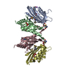
Mass: 22.990 Da / Num. of mol.: 1 / Source method: obtained synthetically / Formula: Na
#3: Chemical
ChemComp-UNL / UNKNOWN LIGAND


Num. of mol.: 4 / Source method: obtained synthetically
#4: Chemical
ChemComp-GOL / GLYCEROL / GLYCERIN / PROPANE-1,2,3-TRIOL


Mass: 92.094 Da / Num. of mol.: 4 / Source method: obtained synthetically / Formula: C3H8O3
#5: Water ChemComp-HOH / water


Mass: 18.015 Da / Num. of mol.: 215 / Source method: isolated from a natural source / Formula: H2O
Sequence detailsSEQUENCE THE CONSTRUCT WAS EXPRESSED WITH A PURIFICATION TAG MGSDKIHHHHHHENLYFQG. THE TAG WAS ...SEQUENCE THE CONSTRUCT WAS EXPRESSED WITH A PURIFICATION TAG MGSDKIHHHHHHENLYFQG. THE TAG WAS REMOVED WITH TEV PROTEASE LEAVING ONLY A GLYCINE (0) FOLLOWED BY THE TARGET SEQUENCE.

-
Experimental details

-
Experiment

ExperimentMethod: X-RAY DIFFRACTION / Number of used crystals: 1

-
Sample preparation

CrystalDensity Matthews: 4.65 Å3/Da / Density % sol: 73.57 %
Crystal growTemperature: 277 K / Method: vapor diffusion, sitting drop / pH: 4.6
Details: 2.0000M NaCl, 0.1M Acetate pH 4., NANODROP, pH 4.6, VAPOR DIFFUSION, SITTING DROP, temperature 277K

-
Data collection

DiffractionMean temperature: 100 K
Diffraction sourceSource: SYNCHROTRON / Site: SSRL / Beamline: BL11-1 / Wavelength: 0.91837,0.97879,0.97932
DetectorType: MARMOSAIC 325 mm CCD / Detector: CCD / Date: Mar 4, 2008 / Details: Flat mirror (vertical focusing)
RadiationMonochromator: Single crystal Si(111) bent monochromator (horizontal focusing)
Protocol: MAD / Monochromatic (M) / Laue (L): M / Scattering type: x-ray
Radiation wavelength
IDWavelength (Å)Relative weight
10.918371
20.978791
30.979321
ReflectionResolution: 2.4→29.841 Å / Num. obs: 42374 / % possible obs: 98.7 % / Redundancy: 3.7 % / Biso Wilson estimate: 54.294 Å2 / Rmerge(I) obs: 0.078 / Rsym value: 0.078 / Net I/σ(I): 6.9
Reflection shell

Diffraction-ID: 1

Resolution (Å)Redundancy (%)Rmerge(I) obsMean I/σ(I) obsNum. measured allNum. unique allRsym value% possible all
2.4-2.463.70.6581.21164131190.65899.8
2.46-2.533.70.5691.31135430520.56999.6
2.53-2.63.70.4631.71119029890.46399.7
2.6-2.683.70.3592.11070528790.35999.6
2.68-2.773.70.2712.81046327950.27199.3
2.77-2.873.70.2243.41016427150.22499.5
2.87-2.983.80.1854.1983826200.18599.1
2.98-3.13.70.1325.7934525000.13299.4
3.1-3.243.70.1017.1911224300.10199
3.24-3.393.80.0848.1864122930.08499.1
3.39-3.583.80.0738.6827221950.07398.8
3.58-3.793.70.0658.6764420390.06598.4
3.79-4.063.80.0629.2744419710.06298.3
4.06-4.383.80.05310.4677117890.05397.7
4.38-4.83.80.04312.2628116600.04397.4
4.8-5.373.80.03713.8572815100.03797.2
5.37-6.23.80.04612.1497713240.04697
6.2-7.593.80.05110.2427311390.05197.1
7.59-10.733.70.03814.732528760.03895.5
10.73-29.843.50.0429.516874790.04290.4

-
Phasing

PhasingMethod: MAD

-
Processing

Software
NameVersionClassificationNB
REFMAC5.2.0019refinement
PHENIXrefinement
SOLVEphasing
MolProbity3beta29model building
SCALAdata scaling
PDB_EXTRACT3.004data extraction
MOSFLMdata reduction
RefinementMethod to determine structure: MAD / Resolution: 2.4→29.841 Å / Cor.coef. Fo:Fc: 0.968 / Cor.coef. Fo:Fc free: 0.95 / SU B: 10.661 / SU ML: 0.129 / TLS residual ADP flag: LIKELY RESIDUAL / Cross valid method: THROUGHOUT / σ(F): 0 / ESU R: 0.195 / ESU R Free: 0.176
Stereochemistry target values: MAXIMUM LIKELIHOOD WITH PHASES
Details: 1. HYDROGENS HAVE BEEN ADDED IN THE RIDING POSITIONS. 2. ATOM RECORD CONTAINS RESIDUAL B FACTORS ONLY. 3. A MET-INHIBITION PROTOCOL WAS USED FOR SELENOMETHIONINE INCORPORATION DURING PROTEIN ...Details: 1. HYDROGENS HAVE BEEN ADDED IN THE RIDING POSITIONS. 2. ATOM RECORD CONTAINS RESIDUAL B FACTORS ONLY. 3. A MET-INHIBITION PROTOCOL WAS USED FOR SELENOMETHIONINE INCORPORATION DURING PROTEIN EXPRESSION. THE OCCUPANCY OF THE SE ATOMS IN THE MSE RESIDUES WAS REDUCED TO 0.75 TO ACCOUNT FOR THE REDUCED SCATTERING POWER DUE TO PARTIAL S-MET INCORPORATION. 4. AN UNKNOWN LIGAND (UNL) WAS MODELED AT THE PUTATIVE ACTIVE SITE ON EACH SUBUNIT IN THE CRYSTALLOGRAPHIC ASYMMETRIC UNIT. 5. GLYCEROL MOLECULES USED AS A CRYOPROTECTANT WERE MODELED INTO THE STRUCTURE. UNEXPLAINED ELECTRON DENSITY AT THE N-TERMINAL REGION OF SUBUNIT B WAS NOT MODELED.
RfactorNum. reflection% reflectionSelection details
Rfree0.219 2140 5.1 %RANDOM
Rwork0.182 ---
obs0.184 42374 98.3 %-
Solvent computationIon probe radii: 0.8 Å / Shrinkage radii: 0.8 Å / VDW probe radii: 1.2 Å / Solvent model: MASK
Displacement parametersBiso mean: 38.537 Å2
Baniso -1Baniso -2Baniso -3
1-1.28 Å20 Å20 Å2
2--0.49 Å20 Å2
3----1.77 Å2
Refinement stepCycle: LAST / Resolution: 2.4→29.841 Å
ProteinNucleic acidLigandSolventTotal
Num. atoms4016 0 52 215 4283
Refine LS restraints
Refine-IDTypeDev idealDev ideal targetNumber
X-RAY DIFFRACTIONr_bond_refined_d0.0170.0224186
X-RAY DIFFRACTIONr_bond_other_d0.0020.022719
X-RAY DIFFRACTIONr_angle_refined_deg1.371.9595719
X-RAY DIFFRACTIONr_angle_other_deg0.90336659
X-RAY DIFFRACTIONr_dihedral_angle_1_deg6.4895552
X-RAY DIFFRACTIONr_dihedral_angle_2_deg30.87824.142169
X-RAY DIFFRACTIONr_dihedral_angle_3_deg14.93415655
X-RAY DIFFRACTIONr_dihedral_angle_4_deg14.4561522
X-RAY DIFFRACTIONr_chiral_restr0.0810.2669
X-RAY DIFFRACTIONr_gen_planes_refined0.0050.024720
X-RAY DIFFRACTIONr_gen_planes_other0.0010.02858
X-RAY DIFFRACTIONr_nbd_refined0.1960.2764
X-RAY DIFFRACTIONr_nbd_other0.1930.22756
X-RAY DIFFRACTIONr_nbtor_refined0.1790.21981
X-RAY DIFFRACTIONr_nbtor_other0.0840.22330
X-RAY DIFFRACTIONr_xyhbond_nbd_refined0.2240.2243
X-RAY DIFFRACTIONr_metal_ion_refined0.1460.23
X-RAY DIFFRACTIONr_symmetry_vdw_refined0.2330.26
X-RAY DIFFRACTIONr_symmetry_vdw_other0.1650.219
X-RAY DIFFRACTIONr_symmetry_hbond_refined0.0330.22
X-RAY DIFFRACTIONr_mcbond_it1.32832676
X-RAY DIFFRACTIONr_mcbond_other0.40831081
X-RAY DIFFRACTIONr_mcangle_it2.45654316
X-RAY DIFFRACTIONr_scbond_it4.25681570
X-RAY DIFFRACTIONr_scangle_it6.057111390
Refine LS restraints NCS

Ens-ID: 1 / Number: 1553 / Refine-ID: X-RAY DIFFRACTION

Dom-IDAuth asym-IDTypeRms dev position (Å)Weight position
1AMEDIUM POSITIONAL0.730.5
2BMEDIUM POSITIONAL0.360.5
3CMEDIUM POSITIONAL0.370.5
4DMEDIUM POSITIONAL0.540.5
1AMEDIUM THERMAL0.672
2BMEDIUM THERMAL0.672
3CMEDIUM THERMAL0.682
4DMEDIUM THERMAL0.732
LS refinement shellResolution: 2.4→2.462 Å / Total num. of bins used: 20
RfactorNum. reflection% reflection
Rfree0.344 156 -
Rwork0.28 2959 -
all-3115 -
obs--99.68 %
Refinement TLS params.

Method: refined / Refine-ID: X-RAY DIFFRACTION

IDL112)L122)L132)L222)L232)L332)S11 (Å °)S12 (Å °)S13 (Å °)S21 (Å °)S22 (Å °)S23 (Å °)S31 (Å °)S32 (Å °)S33 (Å °)T112)T122)T132)T222)T232)T332)Origin x (Å)Origin y (Å)Origin z (Å)
14.20260.50461.24481.84430.34274.00780.17570.2192-0.1044-0.0897-0.06680.20720.0318-0.0804-0.1088-0.0986-0.043-0.0013-0.1551-0.0755-0.204415.871439.4144-23.5833
22.89251.10440.74444.19760.41542.8971-0.18540.11960.3595-0.08740.22360.3303-0.1730.119-0.0382-0.1361-0.0595-0.0606-0.16560.0318-0.166526.578352.1112-10.84
33.214-0.41760.77013.6815-0.05223.3655-0.16-0.08990.41920.03660.1874-0.2283-0.1086-0.0884-0.0274-0.16370.0583-0.0374-0.2021-0.0128-0.164534.809552.296510.9275
45.0706-0.1236-0.07242.5724-0.76133.53580.0985-0.4531-0.00620.0039-0.1541-0.33030.12540.26180.0556-0.1360.08120.011-0.07980.0916-0.203345.697440.239523.5278
Refinement TLS group
IDRefine-IDRefine TLS-IDAuth asym-IDLabel asym-IDAuth seq-IDLabel seq-ID
1X-RAY DIFFRACTION1AA3 - 1344 - 135
2X-RAY DIFFRACTION2BB1 - 1342 - 135
3X-RAY DIFFRACTION3CC1 - 1342 - 135
4X-RAY DIFFRACTION4DD5 - 1346 - 135

+
About Yorodumi

-
News

-
Feb 9, 2022. New format data for meta-information of EMDB entries

New format data for meta-information of EMDB entries

  • Version 3 of the EMDB header file is now the official format.
  • The previous official version 1.9 will be removed from the archive.

Related info.:EMDB header

External links:wwPDB to switch to version 3 of the EMDB data model

-
Aug 12, 2020. Covid-19 info

Covid-19 info

URL: https://pdbjlvh1.pdbj.org/emnavi/covid19.php

New page: Covid-19 featured information page in EM Navigator.

Related info.:Covid-19 info / Mar 5, 2020. Novel coronavirus structure data

+
Mar 5, 2020. Novel coronavirus structure data

Novel coronavirus structure data

Related info.:Yorodumi Speices / Aug 12, 2020. Covid-19 info

External links:COVID-19 featured content - PDBj / Molecule of the Month (242):Coronavirus Proteases

+
Jan 31, 2019. EMDB accession codes are about to change! (news from PDBe EMDB page)

EMDB accession codes are about to change! (news from PDBe EMDB page)

  • The allocation of 4 digits for EMDB accession codes will soon come to an end. Whilst these codes will remain in use, new EMDB accession codes will include an additional digit and will expand incrementally as the available range of codes is exhausted. The current 4-digit format prefixed with “EMD-” (i.e. EMD-XXXX) will advance to a 5-digit format (i.e. EMD-XXXXX), and so on. It is currently estimated that the 4-digit codes will be depleted around Spring 2019, at which point the 5-digit format will come into force.
  • The EM Navigator/Yorodumi systems omit the EMD- prefix.

Related info.:Q: What is EMD? / ID/Accession-code notation in Yorodumi/EM Navigator

External links:EMDB Accession Codes are Changing Soon! / Contact to PDBj

+
Jul 12, 2017. Major update of PDB

Major update of PDB

  • wwPDB released updated PDB data conforming to the new PDBx/mmCIF dictionary.
  • This is a major update changing the version number from 4 to 5, and with Remediation, in which all the entries are updated.
  • In this update, many items about electron microscopy experimental information are reorganized (e.g. em_software).
  • Now, EM Navigator and Yorodumi are based on the updated data.

External links:wwPDB Remediation / Enriched Model Files Conforming to OneDep Data Standards Now Available in the PDB FTP Archive

-
Yorodumi

Thousand views of thousand structures

  • Yorodumi is a browser for structure data from EMDB, PDB, SASBDB, etc.
  • This page is also the successor to EM Navigator detail page, and also detail information page/front-end page for Omokage search.
  • The word "yorodu" (or yorozu) is an old Japanese word meaning "ten thousand". "mi" (miru) is to see.

Related info.:EMDB / PDB / SASBDB / Comparison of 3 databanks / Yorodumi Search / Aug 31, 2016. New EM Navigator & Yorodumi / Yorodumi Papers / Jmol/JSmol / Function and homology information / Changes in new EM Navigator and Yorodumi

Read more