 Movie
Movie Controller

[English] 日本語
 Yorodumi
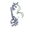
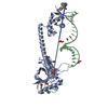
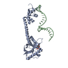
- PDB-3d71: Crystal structure of E253Q BMRR bound to 22 base pair promoter site -
+ Open data

- Basic information
| Entry | Database: PDB / ID: 3d71 | ||||||
|---|---|---|---|---|---|---|---|
| Title | Crystal structure of E253Q BMRR bound to 22 base pair promoter site | ||||||
 Components Components |
| ||||||
 Keywords Keywords | TRANSCRIPTION REGULATOR/DNA / TRANSCRIPTION REGULATOR / PROTEIN-DNA COMPLEX / MULTIDRUG BINDING PROTEIN / MERR FAMILY / WINGED-HELIX / Activator / DNA-binding / Transcription regulation / TRANSCRIPTION REGULATOR-DNA COMPLEX | ||||||
| Function / homology |  Function and homology information Function and homology informationDNA-binding transcription factor activity / regulation of DNA-templated transcription / DNA binding Similarity search - Function | ||||||
| Biological species |  synthetic construct (others) | ||||||
| Method |  X-RAY DIFFRACTION / SYNCHROTRON / MOLECULAR REPLACEMENT / Resolution: 2.8 Å X-RAY DIFFRACTION / SYNCHROTRON / MOLECULAR REPLACEMENT / Resolution: 2.8 Å | ||||||
 Authors Authors | Newberry, K.J. / Huffman, J.L. / Brennan, R.G. | ||||||
 Citation Citation | Journal: J.Biol.Chem. / Year: 2008 Title: Structures of BmrR-Drug Complexes Reveal a Rigid Multidrug Binding Pocket and Transcription Activation through Tyrosine Expulsion Authors: Newberry, K.J. / Huffman, J.L. / Miller, M.C. / Vazquez-Laslop, N. / Neyfakh, A.A. / Brennan, R.G. #1: Journal: J.Biol.Chem. / Year: 2004Title: The Structural Mechanism for Transcription Activation by Merr Family Member Multidrug Transporter Activation, N Terminus. Authors: Newberry, K.J. / Brennan, R.G. | ||||||
| History |
|
- Structure visualization
| Structure viewer | Molecule: MolmilJmol/JSmol |
|---|
- Downloads & links
-Download
| PDBx/mmCIF format | 3d71.cif.gz | 89.5 KB | Display | PDBx/mmCIF format |
|---|---|---|---|---|
| PDB format | pdb3d71.ent.gz | 64.3 KB | Display | PDB format |
| PDBx/mmJSON format | 3d71.json.gz | Tree view | PDBx/mmJSON format | |
| Others |  Other downloads Other downloads |
-Validation report
| Arichive directory | https://data.pdbj.org/pub/pdb/validation_reports/d7/3d71ftp://data.pdbj.org/pub/pdb/validation_reports/d7/3d71 | HTTPS FTP |
|---|
-Related structure data
| Related structure data |  3d6yC  3d6zC  3d70C  1exjS C: citing same article ( S: Starting model for refinement |
|---|---|
| Similar structure data |


-Links
 PDBj
PDBj


- Assembly
| Deposited unit | 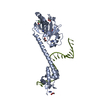
| ||||||||
|---|---|---|---|---|---|---|---|---|---|
| 1 | 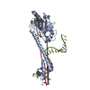
| ||||||||
| Unit cell |
|
-Components
-DNA chain / Protein , 2 types, 2 molecules MA
| #1: DNA chain | Mass: 7362.729 Da / Num. of mol.: 1 / Source method: obtained synthetically / Source: (synth.) synthetic construct (others) |
|---|---|
| #2: Protein | Mass: 33477.219 Da / Num. of mol.: 1 / Fragment: residues 1-278 / Mutation: E253Q, A277L, E278D Source method: isolated from a genetically manipulated source Source: (gene. exp.) |
-Non-polymers , 6 types, 66 molecules 


| #3: Chemical | ChemComp-ZN /  Mass: 65.409 Da / Num. of mol.: 1 / Source method: obtained synthetically / Formula: Zn Mass: 65.409 Da / Num. of mol.: 1 / Source method: obtained synthetically / Formula: Zn | ||||||
|---|---|---|---|---|---|---|---|
| #4: Chemical | ChemComp-FLC /  Mass: 189.100 Da / Num. of mol.: 1 / Source method: obtained synthetically / Formula: C6H5O7 Mass: 189.100 Da / Num. of mol.: 1 / Source method: obtained synthetically / Formula: C6H5O7 | ||||||
| #5: Chemical |  Mass: 76.094 Da / Num. of mol.: 3 / Source method: obtained synthetically / Formula: C3H8O2 Mass: 76.094 Da / Num. of mol.: 3 / Source method: obtained synthetically / Formula: C3H8O2#6: Chemical | ChemComp-ETF / |  Mass: 100.040 Da / Num. of mol.: 1 / Source method: obtained synthetically / Formula: C2H3F3O Mass: 100.040 Da / Num. of mol.: 1 / Source method: obtained synthetically / Formula: C2H3F3O#7: Chemical | ChemComp-IMD /  Mass: 69.085 Da / Num. of mol.: 5 / Source method: obtained synthetically / Formula: C3H5N2 Mass: 69.085 Da / Num. of mol.: 5 / Source method: obtained synthetically / Formula: C3H5N2#8: Water | ChemComp-HOH / | Mass: 18.015 Da / Num. of mol.: 55 / Source method: isolated from a natural source / Formula: H2O |
-Experimental details
-Experiment
| Experiment | Method: X-RAY DIFFRACTION / Number of used crystals: 1 |
|---|
- Sample preparation
| Crystal | Density Matthews: 5.3 Å3/Da / Density % sol: 79 % | ||||||||||||||||||||||||
|---|---|---|---|---|---|---|---|---|---|---|---|---|---|---|---|---|---|---|---|---|---|---|---|---|---|
| Crystal grow | Temperature: 298 K / Method: vapor diffusion, hanging drop / pH: 8 Details: SODIUM CITRATE, IMIDAZOLE, TRIFLUOROETHANOL, pH 8.00, VAPOR DIFFUSION, HANGING DROP, temperature 298K | ||||||||||||||||||||||||
| Components of the solutions |
|
-Data collection
| Diffraction | Mean temperature: 100 K |
|---|---|
| Diffraction source | Source: SYNCHROTRON / Site: SSRL  / Beamline: BL9-1 / Wavelength: 1.08 / Beamline: BL9-1 / Wavelength: 1.08 |
| Detector | Type: MARRESEARCH / Detector: IMAGE PLATE / Date: Nov 1, 2001 / Details: MIRRORS |
| Radiation | Monochromator: SILICON SINGLE CRYSTAL / Protocol: SINGLE WAVELENGTH / Monochromatic (M) / Laue (L): M / Scattering type: x-ray |
| Radiation wavelength | Wavelength: 1.08 Å / Relative weight: 1 |
| Reflection | Resolution: 2.8→47.2 Å / Num. obs: 20900 / % possible obs: 99.3 % / Observed criterion σ(I): 0 / Redundancy: 3.6 % / Biso Wilson estimate: 45.3 Å2 / Rsym value: 0.051 / Net I/σ(I): 10 |
| Reflection shell | Resolution: 2.8→2.86 Å / Redundancy: 3.7 % / Mean I/σ(I) obs: 1.6 / Rsym value: 0.454 / % possible all: 99.9 |
- Processing
| Software |
| ||||||||||||||||||||||||||||||||||||||||||||||||||||||||||||
|---|---|---|---|---|---|---|---|---|---|---|---|---|---|---|---|---|---|---|---|---|---|---|---|---|---|---|---|---|---|---|---|---|---|---|---|---|---|---|---|---|---|---|---|---|---|---|---|---|---|---|---|---|---|---|---|---|---|---|---|---|---|
| Refinement | Method to determine structure: MOLECULAR REPLACEMENT Starting model: PDB entry 1EXJ Resolution: 2.8→47.17 Å / Rfactor Rfree error: 0.008 / Isotropic thermal model: RESTRAINED / Cross valid method: THROUGHOUT / σ(F): 0 / Stereochemistry target values: ENGH & HUBER
| ||||||||||||||||||||||||||||||||||||||||||||||||||||||||||||
| Solvent computation | Solvent model: FLAT MODEL / Bsol: 48.42 Å2 / ksol: 0.34 e/Å3 | ||||||||||||||||||||||||||||||||||||||||||||||||||||||||||||
| Displacement parameters | Biso mean: 65.2 Å2
| ||||||||||||||||||||||||||||||||||||||||||||||||||||||||||||
| Refinement step | Cycle: LAST / Resolution: 2.8→47.17 Å
| ||||||||||||||||||||||||||||||||||||||||||||||||||||||||||||
| Refine LS restraints |
| ||||||||||||||||||||||||||||||||||||||||||||||||||||||||||||
| LS refinement shell | Resolution: 2.8→2.98 Å / Rfactor Rfree error: 0.026 / Total num. of bins used: 6
| ||||||||||||||||||||||||||||||||||||||||||||||||||||||||||||
| Xplor file |
|
