 Movie
Movie Controller

[English] 日本語
 Yorodumi
- PDB-3cyv: Crystal structure of uroporphyrinogen decarboxylase from Shigella... -
+ Open data

- Basic information
| Entry | Database: PDB / ID: 3cyv | ||||||
|---|---|---|---|---|---|---|---|
| Title | Crystal structure of uroporphyrinogen decarboxylase from Shigella flexineri: new insights into its catalytic mechanism | ||||||
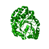
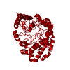
 Components Components | Uroporphyrinogen decarboxylase | ||||||
 Keywords Keywords | LYASE / alpha/beta barrel / Cytoplasm / Decarboxylase / Porphyrin biosynthesis | ||||||
| Function / homology |  Function and homology information Function and homology information: / uroporphyrinogen decarboxylase / uroporphyrinogen decarboxylase activity / cytosol Similarity search - Function | ||||||
| Biological species |  Shigella flexneri (bacteria) Shigella flexneri (bacteria) | ||||||
| Method |  X-RAY DIFFRACTION / SYNCHROTRON / MOLECULAR REPLACEMENT / Resolution: 2.8 Å X-RAY DIFFRACTION / SYNCHROTRON / MOLECULAR REPLACEMENT / Resolution: 2.8 Å | ||||||
 Authors Authors | Liu, H. / Zhou, H. / Bi, R. | ||||||
 Citation Citation | Journal: To be Published Title: Crystal structure of uroporphyrinogen III decarboxylase from Shigella flexineri: new insights into its catalytic mechanism Authors: Liu, H. / Zhou, H. / Bi, R. | ||||||
| History |
|
- Structure visualization
| Structure viewer | Molecule: MolmilJmol/JSmol |
|---|
- Downloads & links
-Download
| PDBx/mmCIF format | 3cyv.cif.gz | 84.2 KB | Display | PDBx/mmCIF format |
|---|---|---|---|---|
| PDB format | pdb3cyv.ent.gz | 63.3 KB | Display | PDB format |
| PDBx/mmJSON format | 3cyv.json.gz | Tree view | PDBx/mmJSON format | |
| Others |  Other downloads Other downloads |
-Validation report
| Arichive directory | https://data.pdbj.org/pub/pdb/validation_reports/cy/3cyvftp://data.pdbj.org/pub/pdb/validation_reports/cy/3cyv | HTTPS FTP |
|---|
-Related structure data
| Related structure data |  1uroS S: Starting model for refinement |
|---|---|
| Similar structure data |


-Links
 PDBj
PDBj- Assembly
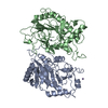
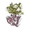
| Deposited unit | 
| ||||||||
|---|---|---|---|---|---|---|---|---|---|
| 1 | 
| ||||||||
| Unit cell |
|
-Components
| #1: Protein | Mass: 39274.988 Da / Num. of mol.: 1 Source method: isolated from a genetically manipulated source Source: (gene. exp.) Shigella flexneri (bacteria) / Strain: 2a str. 301 / Gene: urod / Plasmid: pET22b(+) / Production host: |
|---|---|
| #2: Water | ChemComp-HOH /  Mass: 18.015 Da / Num. of mol.: 229 / Source method: isolated from a natural source / Formula: H2O Mass: 18.015 Da / Num. of mol.: 229 / Source method: isolated from a natural source / Formula: H2O |
-Experimental details
-Experiment
| Experiment | Method: X-RAY DIFFRACTION / Number of used crystals: 1 |
|---|
- Sample preparation
| Crystal | Density Matthews: 2.31 Å3/Da / Density % sol: 46.7 % |
|---|
-Data collection
| Diffraction | Mean temperature: 100 K |
|---|---|
| Diffraction source | Source: SYNCHROTRON / Site: BSRF  / Beamline: 3W1A / Wavelength: 0.97954 Å / Beamline: 3W1A / Wavelength: 0.97954 Å |
| Detector | Type: MAR CCD 165 mm / Detector: CCD / Date: Mar 26, 2005 |
| Radiation | Monochromator: SI(111) DOUBLE-CRYSTAL / Protocol: SINGLE WAVELENGTH / Monochromatic (M) / Laue (L): M / Scattering type: x-ray |
| Radiation wavelength | Wavelength: 0.97954 Å / Relative weight: 1 |
| Reflection | Resolution: 2.8→50 Å / Num. all: 9336 / Num. obs: 9240 / % possible obs: 99.3 % / Observed criterion σ(F): 0 / Observed criterion σ(I): 0 / Redundancy: 6.7 % / Biso Wilson estimate: 31.1 Å2 / Rmerge(I) obs: 0.113 / Net I/σ(I): 17.9 |
| Reflection shell | Resolution: 2.8→2.9 Å / Redundancy: 6.4 % / Rmerge(I) obs: 0.309 / Mean I/σ(I) obs: 6 / Num. unique all: 853 / % possible all: 92.7 |
- Processing
| Software |
| ||||||||||||||||||||||||||||||||||||||||||||||||||||||||||||
|---|---|---|---|---|---|---|---|---|---|---|---|---|---|---|---|---|---|---|---|---|---|---|---|---|---|---|---|---|---|---|---|---|---|---|---|---|---|---|---|---|---|---|---|---|---|---|---|---|---|---|---|---|---|---|---|---|---|---|---|---|---|
| Refinement | Method to determine structure: MOLECULAR REPLACEMENT Starting model: PDB ENTRY 1URO Resolution: 2.8→50 Å / Rfactor Rfree error: 0.012 / Data cutoff high absF: 1764582.51 / Data cutoff low absF: 0 / Isotropic thermal model: anisotropic / Cross valid method: THROUGHOUT / σ(F): 0 / Stereochemistry target values: Engh & Huber
| ||||||||||||||||||||||||||||||||||||||||||||||||||||||||||||
| Solvent computation | Solvent model: FLAT MODEL / Bsol: 33.2071 Å2 / ksol: 0.337117 e/Å3 | ||||||||||||||||||||||||||||||||||||||||||||||||||||||||||||
| Displacement parameters | Biso mean: 23.9 Å2
| ||||||||||||||||||||||||||||||||||||||||||||||||||||||||||||
| Refine analyze |
| ||||||||||||||||||||||||||||||||||||||||||||||||||||||||||||
| Refinement step | Cycle: LAST / Resolution: 2.8→50 Å
| ||||||||||||||||||||||||||||||||||||||||||||||||||||||||||||
| Refine LS restraints |
| ||||||||||||||||||||||||||||||||||||||||||||||||||||||||||||
| LS refinement shell | Resolution: 2.8→2.98 Å / Rfactor Rfree error: 0.036 / Total num. of bins used: 6
| ||||||||||||||||||||||||||||||||||||||||||||||||||||||||||||
| Xplor file |
|
