 Movie
Movie Controller


+ Open data

- Basic information
| Entry | Database: PDB / ID: 3cxs | ||||||
|---|---|---|---|---|---|---|---|
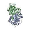
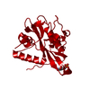
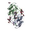
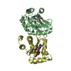
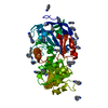
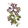
| Title | Crystal structure of human GNA1 | ||||||
 Components Components | Glucosamine 6-phosphate N-acetyltransferase | ||||||
 Keywords Keywords | TRANSFERASE / Glucosamine-6-phosphate N-acetyltransferase 1 / Acyltransferase / Endosome / Golgi apparatus / Membrane | ||||||
| Function / homology |  Function and homology information Function and homology informationglucosamine-phosphate N-acetyltransferase / glucosamine 6-phosphate N-acetyltransferase activity / Synthesis of UDP-N-acetyl-glucosamine / UDP-N-acetylglucosamine biosynthetic process / endoplasmic reticulum-Golgi intermediate compartment / cellular response to leukemia inhibitory factor / late endosome / endosome membrane / Golgi membrane / identical protein binding / cytosol Similarity search - Function | ||||||
| Biological species |  Homo sapiens (human) Homo sapiens (human) | ||||||
| Method |  X-RAY DIFFRACTION / MOLECULAR REPLACEMENT / molecular replacement / Resolution: 2.7 Å X-RAY DIFFRACTION / MOLECULAR REPLACEMENT / molecular replacement / Resolution: 2.7 Å | ||||||
 Authors Authors | Wang, J. / Liu, X. / Li, L.-F. / Su, X.-D. | ||||||
 Citation Citation | Journal: Febs Lett. / Year: 2008 Title: Acceptor substrate binding revealed by crystal structure of human glucosamine-6-phosphate N-acetyltransferase 1 Authors: Wang, J. / Liu, X. / Liang, Y.-H. / Li, L.-F. / Su, X.-D. | ||||||
| History |
|
- Structure visualization
| Structure viewer | Molecule: MolmilJmol/JSmol |
|---|
- Downloads & links
-Download
| PDBx/mmCIF format | 3cxs.cif.gz | 48.1 KB | Display | PDBx/mmCIF format |
|---|---|---|---|---|
| PDB format | pdb3cxs.ent.gz | 33.5 KB | Display | PDB format |
| PDBx/mmJSON format | 3cxs.json.gz | Tree view | PDBx/mmJSON format | |
| Others |  Other downloads Other downloads |
-Validation report
| Summary document | 3cxs_validation.pdf.gz | 421.2 KB | Display | wwPDB validaton report |
|---|---|---|---|---|
| Full document | 3cxs_full_validation.pdf.gz | 424.4 KB | Display | |
| Data in XML | 3cxs_validation.xml.gz | 8.8 KB | Display | |
| Data in CIF | 3cxs_validation.cif.gz | 10.8 KB | Display | |
| Arichive directory | https://data.pdbj.org/pub/pdb/validation_reports/cx/3cxsftp://data.pdbj.org/pub/pdb/validation_reports/cx/3cxs | HTTPS FTP |
-Related structure data
| Related structure data |  3cxpC  3cxqC  2huzS C: citing same article ( S: Starting model for refinement |
|---|---|
| Similar structure data |


-Links
 PDBj
PDBj

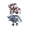
- Assembly
| Deposited unit | 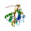
| ||||||||
|---|---|---|---|---|---|---|---|---|---|
| 1 | 
| ||||||||
| Unit cell |
|
-Components
| #1: Protein | Mass: 20776.117 Da / Num. of mol.: 1 Source method: isolated from a genetically manipulated source Source: (gene. exp.) Homo sapiens (human) / Gene: GNPNAT1 / Plasmid: pET28a / Production host:  References: UniProt: Q96EK6, glucosamine-phosphate N-acetyltransferase |
|---|---|
| #2: Water | ChemComp-HOH /  Mass: 18.015 Da / Num. of mol.: 3 / Source method: isolated from a natural source / Formula: H2O Mass: 18.015 Da / Num. of mol.: 3 / Source method: isolated from a natural source / Formula: H2O |
-Experimental details
-Experiment
| Experiment | Method: X-RAY DIFFRACTION / Number of used crystals: 1 |
|---|
- Sample preparation
| Crystal | Density Matthews: 2.12 Å3/Da / Density % sol: 42.96 % |
|---|---|
| Crystal grow | Temperature: 289 K / Method: vapor diffusion, hanging drop / pH: 6.3 Details: 0.2M MgCl2, 0.1M Bis-Tris pH 6.3, 25% w/v PEG 3350, vapor diffusion, hanging drop, temperature 289K |
-Data collection
| Diffraction | Mean temperature: 100 K | ||||||||||||||||||||||||||||||||||||
|---|---|---|---|---|---|---|---|---|---|---|---|---|---|---|---|---|---|---|---|---|---|---|---|---|---|---|---|---|---|---|---|---|---|---|---|---|---|
| Diffraction source | Source: ROTATING ANODE / Type: BRUKER AXS MICROSTAR-H / Wavelength: 1.5418 Å | ||||||||||||||||||||||||||||||||||||
| Detector | Type: BRUKER SMART 6000 / Detector: CCD / Date: Jan 15, 2008 / Details: Montel mirrors | ||||||||||||||||||||||||||||||||||||
| Radiation | Monochromator: Montel mirrors / Protocol: SINGLE WAVELENGTH / Monochromatic (M) / Laue (L): M / Scattering type: x-ray | ||||||||||||||||||||||||||||||||||||
| Radiation wavelength | Wavelength: 1.5418 Å / Relative weight: 1 | ||||||||||||||||||||||||||||||||||||
| Reflection | Resolution: 2.7→50 Å / Num. all: 5481 / Num. obs: 5121 / % possible obs: 94.49 % / Redundancy: 5.53 % / Rsym value: 0.063 / Χ2: 0.73 / Net I/σ(I): 8.96 / Scaling rejects: 1374 | ||||||||||||||||||||||||||||||||||||
| Reflection shell |
|
-Phasing
| Phasing | Method: molecular replacement | |||||||||
|---|---|---|---|---|---|---|---|---|---|---|
| Phasing MR |
|
- Processing
| Software |
| ||||||||||||||||||||||||||||||||||||||||||||||||||||||||||||||||||||||||||||||||||||||||||
|---|---|---|---|---|---|---|---|---|---|---|---|---|---|---|---|---|---|---|---|---|---|---|---|---|---|---|---|---|---|---|---|---|---|---|---|---|---|---|---|---|---|---|---|---|---|---|---|---|---|---|---|---|---|---|---|---|---|---|---|---|---|---|---|---|---|---|---|---|---|---|---|---|---|---|---|---|---|---|---|---|---|---|---|---|---|---|---|---|---|---|---|
| Refinement | Method to determine structure: MOLECULAR REPLACEMENT Starting model: PDB ENTRY 2HUZ Resolution: 2.7→20 Å / Cor.coef. Fo:Fc: 0.897 / Cor.coef. Fo:Fc free: 0.86 / SU B: 17.374 / SU ML: 0.347 / Cross valid method: THROUGHOUT / σ(F): 0 / ESU R Free: 0.428 / Stereochemistry target values: MAXIMUM LIKELIHOOD
| ||||||||||||||||||||||||||||||||||||||||||||||||||||||||||||||||||||||||||||||||||||||||||
| Solvent computation | Ion probe radii: 0.8 Å / Shrinkage radii: 0.8 Å / VDW probe radii: 1.2 Å / Solvent model: MASK | ||||||||||||||||||||||||||||||||||||||||||||||||||||||||||||||||||||||||||||||||||||||||||
| Displacement parameters | Biso mean: 35.282 Å2
| ||||||||||||||||||||||||||||||||||||||||||||||||||||||||||||||||||||||||||||||||||||||||||
| Refinement step | Cycle: LAST / Resolution: 2.7→20 Å
| ||||||||||||||||||||||||||||||||||||||||||||||||||||||||||||||||||||||||||||||||||||||||||
| Refine LS restraints |
| ||||||||||||||||||||||||||||||||||||||||||||||||||||||||||||||||||||||||||||||||||||||||||
| LS refinement shell | Resolution: 2.7→2.769 Å / Total num. of bins used: 20
|
