 Movie
Movie Controller


+ Open data

- Basic information
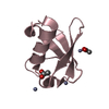
| Entry | Database: PDB / ID: 3cu4 | ||||||
|---|---|---|---|---|---|---|---|
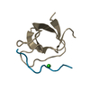
| Title | OmcF, Outer membrance cytochrome F from Geobacter sulfurreducens | ||||||
 Components Components | Cytochrome c family protein | ||||||
 Keywords Keywords | ELECTRON TRANSPORT / cytochrome c6 / Geobacter sulfurreducens / monoheme cytochrome | ||||||
| Function / homology |  Function and homology information Function and homology informationplasma membrane-derived thylakoid lumen / electron transfer activity / iron ion binding / heme binding Similarity search - Function | ||||||
| Biological species |  Geobacter sulfurreducens (bacteria) Geobacter sulfurreducens (bacteria) | ||||||
| Method |  X-RAY DIFFRACTION / SYNCHROTRON / SAD / Resolution: 1.3 Å X-RAY DIFFRACTION / SYNCHROTRON / SAD / Resolution: 1.3 Å | ||||||
 Authors Authors | Pokkuluri, P.R. / Schiffer, M. | ||||||
 Citation Citation | Journal: Proteins / Year: 2008 Title: Outer membrane cytochrome c, OmcF, from Geobacter sulfurreducens: High structural similarity to an algal cytochrome c(6). Authors: Pokkuluri, P.R. / Londer, Y.Y. / Wood, S.J. / Duke, N.E. / Morgado, L. / Salgueiro, C.A. / Schiffer, M. | ||||||
| History |
|
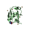
- Structure visualization
| Structure viewer | Molecule: MolmilJmol/JSmol |
|---|
- Downloads & links
-Download
| PDBx/mmCIF format | 3cu4.cif.gz | 31.4 KB | Display | PDBx/mmCIF format |
|---|---|---|---|---|
| PDB format | pdb3cu4.ent.gz | 19.9 KB | Display | PDB format |
| PDBx/mmJSON format | 3cu4.json.gz | Tree view | PDBx/mmJSON format | |
| Others |  Other downloads Other downloads |
-Validation report
| Arichive directory | https://data.pdbj.org/pub/pdb/validation_reports/cu/3cu4ftp://data.pdbj.org/pub/pdb/validation_reports/cu/3cu4 | HTTPS FTP |
|---|
-Related structure data
| Related structure data | |
|---|---|
| Similar structure data |


-Links
 PDBj
PDBj


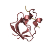
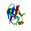
- Assembly
| Deposited unit | 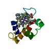
| ||||||||
|---|---|---|---|---|---|---|---|---|---|
| 1 |
| ||||||||
| Unit cell |
|
-Components
| #1: Protein | Mass: 8669.767 Da / Num. of mol.: 1 Source method: isolated from a genetically manipulated source Source: (gene. exp.) Geobacter sulfurreducens (bacteria) / Gene: GSU2432 / Production host: Strain (production host): BL21 (DE3) cotransformed with plasmid pEC86 References: UniProt: Q74AE4 |
|---|---|
| #2: Chemical | ChemComp-HEM /   Mass: 616.487 Da / Num. of mol.: 1 / Source method: obtained synthetically / Formula: C34H32FeN4O4 Mass: 616.487 Da / Num. of mol.: 1 / Source method: obtained synthetically / Formula: C34H32FeN4O4 |
| #3: Water | ChemComp-HOH /  Mass: 18.015 Da / Num. of mol.: 116 / Source method: isolated from a natural source / Formula: H2O Mass: 18.015 Da / Num. of mol.: 116 / Source method: isolated from a natural source / Formula: H2O |
-Experimental details
-Experiment
| Experiment | Method: X-RAY DIFFRACTION / Number of used crystals: 1 |
|---|
- Sample preparation
| Crystal | Density Matthews: 2.14 Å3/Da / Density % sol: 42.44 % |
|---|---|
| Crystal grow | Temperature: 298 K / Method: vapor diffusion / pH: 5 Details: 1.2 M trisodium citrate, 0.1 M Tris pH 8.5 (Salt Rx screen-22), microseeding, pH 5.0, VAPOR DIFFUSION, temperature 298K |
-Data collection
| Diffraction | Mean temperature: 100 K |
|---|---|
| Diffraction source | Source: SYNCHROTRON / Site: APS  / Beamline: 19-BM / Wavelength: 0.97911 Å / Beamline: 19-BM / Wavelength: 0.97911 Å |
| Detector | Type: CUSTOM-MADE / Detector: CCD / Date: Jul 13, 2007 |
| Radiation | Protocol: SINGLE WAVELENGTH / Monochromatic (M) / Laue (L): M / Scattering type: x-ray |
| Radiation wavelength | Wavelength: 0.97911 Å / Relative weight: 1 |
| Reflection | Resolution: 1.3→50 Å / Num. all: 33541 / Num. obs: 33541 / % possible obs: 96 % / Redundancy: 4.2 % / Biso Wilson estimate: 5.9 Å2 / Rmerge(I) obs: 0.037 / Net I/σ(I): 42.4 |
| Reflection shell | Resolution: 1.3→1.31 Å / Redundancy: 2.7 % / Rmerge(I) obs: 0.105 / Mean I/σ(I) obs: 9.7 / Num. unique all: 617 / % possible all: 70 |
- Processing
| Software |
| ||||||||||||||||||||||||||||||||||||
|---|---|---|---|---|---|---|---|---|---|---|---|---|---|---|---|---|---|---|---|---|---|---|---|---|---|---|---|---|---|---|---|---|---|---|---|---|---|
| Refinement | Method to determine structure: SAD / Resolution: 1.3→24.01 Å / Rfactor Rfree error: 0.003 / Data cutoff high absF: 134653.74 / Data cutoff low absF: 0 / Isotropic thermal model: RESTRAINED / Cross valid method: THROUGHOUT / σ(F): 0 / Stereochemistry target values: Engh & Huber Details: THE FOLLOWING SIDE CHAIN ATOMS HAVE WEAK DENSITY AND ARE PROBABLY DISORDERED: HIS47:CD2,ND1,CE1,NE2 GLU49:CD,OE1,OE2 ARG56:NE,CZ,NH1,NH2 LYS94:CE,NZ GLU97:CD,OE1,OE2
| ||||||||||||||||||||||||||||||||||||
| Solvent computation | Solvent model: FLAT MODEL / Bsol: 47.2274 Å2 / ksol: 0.413536 e/Å3 | ||||||||||||||||||||||||||||||||||||
| Displacement parameters | Biso mean: 11 Å2
| ||||||||||||||||||||||||||||||||||||
| Refine analyze |
| ||||||||||||||||||||||||||||||||||||
| Refinement step | Cycle: LAST / Resolution: 1.3→24.01 Å
| ||||||||||||||||||||||||||||||||||||
| Refine LS restraints |
| ||||||||||||||||||||||||||||||||||||
| LS refinement shell | Resolution: 1.3→1.38 Å / Rfactor Rfree error: 0.008 / Total num. of bins used: 6
| ||||||||||||||||||||||||||||||||||||
| Xplor file |
|
