[English] 日本語
Yorodumi
- PDB-3ct9: Crystal structure of a putative zinc peptidase (NP_812461.1) from... -

+
Open data


ID or keywords:

Loading...

-
Basic information

Entry
Database: PDB / ID: 3ct9
TitleCrystal structure of a putative zinc peptidase (NP_812461.1) from Bacteroides thetaiotaomicron VPI-5482 at 2.31 A resolution
ComponentsAcetylornithine deacetylase
KeywordsHYDROLASE / NP_812461.1 / A Putative Zinc Peptidase / Peptidase family M20/M25/M40 / Structural Genomics / Joint Center for Structural Genomics / JCSG / Protein Structure Initiative / PSI-2
Function / homology
Function and homology information


acetylornithine deacetylase activity / arginine biosynthetic process
Similarity search - Function
ArgE / dapE / ACY1 / CPG2 / yscS family signature 1. / ArgE/DapE/ACY1/CPG2/YscS, conserved site / Alpha-Beta Plaits - #360 / Peptidase M20, dimerisation domain / Bacterial exopeptidase dimerisation domain / Peptidase dimerisation domain / Peptidase M20 / Peptidase family M20/M25/M40 / Zn peptidases / Aminopeptidase ...ArgE / dapE / ACY1 / CPG2 / yscS family signature 1. / ArgE/DapE/ACY1/CPG2/YscS, conserved site / Alpha-Beta Plaits - #360 / Peptidase M20, dimerisation domain / Bacterial exopeptidase dimerisation domain / Peptidase dimerisation domain / Peptidase M20 / Peptidase family M20/M25/M40 / Zn peptidases / Aminopeptidase / Alpha-Beta Plaits / 2-Layer Sandwich / 3-Layer(aba) Sandwich / Alpha Beta
Similarity search - Domain/homology
IODIDE ION / DI(HYDROXYETHYL)ETHER / Acetylornithine deacetylase
Similarity search - Component
Biological speciesBacteroides thetaiotaomicron VPI-5482 (bacteria)
MethodX-RAY DIFFRACTION / SYNCHROTRON / SAD / Resolution: 2.31 Å
AuthorsJoint Center for Structural Genomics (JCSG)
CitationJournal: To be published
Title: Crystal structure of a Putative Zinc Peptidase (NP_812461.1) from Bacteroides thetaiotaomicron VPI-5482 at 2.31 A resolution
Authors: Joint Center for Structural Genomics (JCSG)
History
DepositionApr 11, 2008Deposition site: RCSB / Processing site: RCSB
Revision 1.0May 6, 2008Provider: repository / Type: Initial release
Revision 1.1Jul 13, 2011Group: Advisory / Version format compliance
Revision 1.2Oct 25, 2017Group: Author supporting evidence / Refinement description / Category: pdbx_struct_assembly_auth_evidence / software / Item: _software.classification / _software.name
Revision 1.3Jul 24, 2019Group: Data collection / Derived calculations / Refinement description
Category: software / struct_conn
Item: _software.classification / _software.contact_author ..._software.classification / _software.contact_author / _software.contact_author_email / _software.language / _software.location / _software.name / _software.type / _software.version / _struct_conn.pdbx_leaving_atom_flag
Revision 1.4Feb 1, 2023Group: Database references / Derived calculations / Category: database_2 / struct_ref_seq_dif / struct_site
Item: _database_2.pdbx_DOI / _database_2.pdbx_database_accession ..._database_2.pdbx_DOI / _database_2.pdbx_database_accession / _struct_ref_seq_dif.details / _struct_site.pdbx_auth_asym_id / _struct_site.pdbx_auth_comp_id / _struct_site.pdbx_auth_seq_id

-
Structure visualization

Structure viewerMolecule:
MolmilJmol/JSmol

Downloads & links

-
Assembly

Deposited unit
A: Acetylornithine deacetylase
B: Acetylornithine deacetylase
hetero molecules


Theoretical massNumber of molelcules
Total (without water)81,22222
Polymers79,5782
Non-polymers1,64420
Water3,459192
1


  • Idetical with deposited unit
  • defined by author&software
  • Evidence: gel filtration
TypeNameSymmetry operationNumber
identity operation1_555x,y,z1
Buried area2770 Å2
ΔGint-14.3 kcal/mol
Surface area29470 Å2
MethodPISA
Unit cell
Length a, b, c (Å)51.340, 67.250, 107.890
Angle α, β, γ (deg.)90.000, 94.280, 90.000
Int Tables number4
Space group name H-MP1211
Noncrystallographic symmetry (NCS)NCS domain:
IDEns-IDDetails
11A
21B
31A
41B
51A
61B

NCS domain segments:

Ens-ID: 1

Dom-IDComponent-IDBeg label comp-IDEnd label comp-IDRefine codeAuth asym-IDLabel asym-IDAuth seq-IDLabel seq-ID
11TYRLEU5AA3 - 1744 - 175
21TYRLEU5BB3 - 1744 - 175
32MSEASN6AA175 - 279176 - 280
42MSEASN6BB175 - 279176 - 280
53SERLEU5AA280 - 355281 - 356
63SERLEU5BB280 - 355281 - 356
Details
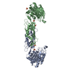
THIS ENTRY CONTAINS THE CRYSTALLOGRAPHIC ASYMMETRIC UNIT WHICH CONSISTS OF 2 CHAINS WHICH FORM A DIMER BASED ON CRYSTAL PACKING ANALYSIS. SIZE EXCLUSION CHROMATOGRAPHY SUPPORTS THE ASSIGNMENT OF A DIMER AS THE SIGNIFICANT OLIGOMERIZATION STATE.

-
Components

-
Protein , 1 types, 2 molecules AB

#1: Protein Acetylornithine deacetylase /


Mass: 39788.844 Da / Num. of mol.: 2
Source method: isolated from a genetically manipulated source
Source: (gene. exp.) Bacteroides thetaiotaomicron VPI-5482 (bacteria)
Species: Bacteroides thetaiotaomicron / Strain: VPI-5482 / DSM 2079 / NCTC 10582 / E50 / Gene: NP_812461.1, BT_3549 / Plasmid: SpeedET / Production host: Escherichia coli (E. coli) / Strain (production host): HK100 / References: UniProt: Q8A1V9

-
Non-polymers , 5 types, 212 molecules

#2: Chemical
ChemComp-IOD / IODIDE ION / Iodide


Mass: 126.904 Da / Num. of mol.: 8 / Source method: obtained synthetically / Formula: I
#3: Chemical
ChemComp-CL / CHLORIDE ION / Chloride


Mass: 35.453 Da / Num. of mol.: 6 / Source method: obtained synthetically / Formula: Cl
#4: Chemical
ChemComp-EDO / 1,2-ETHANEDIOL / ETHYLENE GLYCOL / Ethylene glycol


Mass: 62.068 Da / Num. of mol.: 5 / Source method: obtained synthetically / Formula: C2H6O2
#5: Chemical ChemComp-PEG / DI(HYDROXYETHYL)ETHER / Diethylene glycol


Mass: 106.120 Da / Num. of mol.: 1 / Source method: obtained synthetically / Formula: C4H10O3
#6: Water ChemComp-HOH / water / Water


Mass: 18.015 Da / Num. of mol.: 192 / Source method: isolated from a natural source / Formula: H2O

-
Details

Sequence detailsTHE CONSTRUCT WAS EXPRESSED WITH A PURIFICATION TAG MGSDKIHHHHHHENLYFQG. THE TAG WAS REMOVED WITH ...THE CONSTRUCT WAS EXPRESSED WITH A PURIFICATION TAG MGSDKIHHHHHHENLYFQG. THE TAG WAS REMOVED WITH TEV PROTEASE LEAVING ONLY A GLYCINE FOLLOWED BY THE TARGET SEQUENCE.

-
Experimental details

-
Experiment

ExperimentMethod: X-RAY DIFFRACTION / Number of used crystals: 1

-
Sample preparation

CrystalDensity Matthews: 2.33 Å3/Da / Density % sol: 47.3 %
Crystal growTemperature: 277 K / Method: vapor diffusion, sitting drop / pH: 6.2
Details: NANODROP, 0.2M NH4I, 20.0% PEG 3350, No Buffer pH 6.2, VAPOR DIFFUSION, SITTING DROP, temperature 277K

-
Data collection

DiffractionMean temperature: 100 K
Diffraction sourceSource: SYNCHROTRON / Site: SSRL / Beamline: BL11-1 / Wavelength: 0.9791 Å
DetectorType: MARMOSAIC 325 mm CCD / Detector: CCD / Date: Jun 21, 2007 / Details: Flat mirror (vertical focusing)
RadiationMonochromator: Single crystal Si(111) bent (horizontal focusing)
Protocol: SINGLE WAVELENGTH / Monochromatic (M) / Laue (L): M / Scattering type: x-ray
Radiation wavelengthWavelength: 0.9791 Å / Relative weight: 1
ReflectionResolution: 2.28→28.513 Å / Num. obs: 31807 / % possible obs: 93.7 % / Observed criterion σ(I): -3 / Biso Wilson estimate: 39.305 Å2 / Rmerge(I) obs: 0.043 / Net I/σ(I): 12.23
Reflection shell
Resolution (Å)Rmerge(I) obsMean I/σ(I) obsNum. measured obsNum. unique obsDiffraction-ID% possible all
2.28-2.360.24338802772142.6
2.36-2.460.2093.7117756904199.9
2.46-2.570.1734.5110976475199.8
2.57-2.70.1435.5108366266199.8
2.7-2.870.1067.2113356559199.7
2.87-3.090.0759.9113346496199.7
3.09-3.40.04914.1115926609199.8
3.4-3.890.03618.8115386528199.3
3.89-4.890.02524.2116106486199.1
4.89-28.5130.02126.7116266430196.2

-
Phasing

PhasingMethod: SAD

-
Processing

Software
NameVersionClassificationNB
REFMAC5.2.0019refinement
PHENIXrefinement
SOLVEphasing
MolProbity3beta29model building
XSCALEdata scaling
PDB_EXTRACT3.004data extraction
MAR345CCDdata collection
XDSdata reduction
RefinementMethod to determine structure: SAD / Resolution: 2.31→28.513 Å / Cor.coef. Fo:Fc: 0.944 / Cor.coef. Fo:Fc free: 0.907 / SU B: 14.21 / SU ML: 0.192 / TLS residual ADP flag: LIKELY RESIDUAL / Cross valid method: THROUGHOUT / σ(F): 0 / ESU R: 0.373 / ESU R Free: 0.255
Stereochemistry target values: MAXIMUM LIKELIHOOD WITH PHASES
Details: 1. HYDROGENS HAVE BEEN ADDED IN THE RIDING POSITIONS. 2. ATOM RECORD CONTAINS RESIDUAL B FACTORS ONLY. 3. A MET-INHIBITION PROTOCOL WAS USED FOR SELENOMETHIONINE INCORPORATION DURING PROTEIN ...Details: 1. HYDROGENS HAVE BEEN ADDED IN THE RIDING POSITIONS. 2. ATOM RECORD CONTAINS RESIDUAL B FACTORS ONLY. 3. A MET-INHIBITION PROTOCOL WAS USED FOR SELENOMETHIONINE INCORPORATION DURING PROTEIN EXPRESSION. THE OCCUPANCY OF THE SE ATOMS IN THE MSE RESIDUES WAS REDUCED TO 0.75 FOR THE REDUCED SCATTERING POWER DUE TO PARTIAL S-MET INCORPORATION. 4. IODIDE IONS WERE MODELED BASED ON THE CRYSTALLIZATION CONDITIONS AND ANOMALOUS DIFFERENCE FOURIER PEAKS. 5. CHLORIDE, 1,2-ETHANE DIOL AND PEG WERE MODELED BASED ON CRYSTALLIZATION AND CRYOPROTECTION CONDITIONS. 6. THE CIS PEPTIDE BONDS BETWEEN 104-105 ARE SUPPORTED BY DENSITY. 7. AMINO ACID PRO 87 IN CHAIN B IS A RAMACHANDRAN OUTLIER IN A REGION OF ELECTRON DENSITY THAT IS DIFFICULT TO MODEL.
RfactorNum. reflection% reflectionSelection details
Rfree0.253 1613 5.1 %RANDOM
Rwork0.194 ---
obs0.197 31794 98.43 %-
Solvent computationIon probe radii: 0.8 Å / Shrinkage radii: 0.8 Å / VDW probe radii: 1.2 Å / Solvent model: MASK
Displacement parametersBiso mean: 29.051 Å2
Baniso -1Baniso -2Baniso -3
1--1.27 Å20 Å20.79 Å2
2--0.31 Å20 Å2
3---1.07 Å2
Refinement stepCycle: LAST / Resolution: 2.31→28.513 Å
ProteinNucleic acidLigandSolventTotal
Num. atoms5230 0 41 192 5463
Refine LS restraints
Refine-IDTypeDev idealDev ideal targetNumber
X-RAY DIFFRACTIONr_bond_refined_d0.0160.0225363
X-RAY DIFFRACTIONr_bond_other_d0.0010.023610
X-RAY DIFFRACTIONr_angle_refined_deg1.7321.9797266
X-RAY DIFFRACTIONr_angle_other_deg1.0338847
X-RAY DIFFRACTIONr_dihedral_angle_1_deg3.8985694
X-RAY DIFFRACTIONr_dihedral_angle_2_deg34.63624.491216
X-RAY DIFFRACTIONr_dihedral_angle_3_deg13.10515895
X-RAY DIFFRACTIONr_dihedral_angle_4_deg15.6221530
X-RAY DIFFRACTIONr_chiral_restr0.1070.2837
X-RAY DIFFRACTIONr_gen_planes_refined0.0050.025979
X-RAY DIFFRACTIONr_gen_planes_other0.0010.021015
X-RAY DIFFRACTIONr_nbd_refined0.2020.2926
X-RAY DIFFRACTIONr_nbd_other0.1870.23457
X-RAY DIFFRACTIONr_nbtor_refined0.1680.22488
X-RAY DIFFRACTIONr_nbtor_other0.0880.22636
X-RAY DIFFRACTIONr_xyhbond_nbd_refined0.1850.2168
X-RAY DIFFRACTIONr_symmetry_vdw_refined0.30.29
X-RAY DIFFRACTIONr_symmetry_vdw_other0.2580.219
X-RAY DIFFRACTIONr_symmetry_hbond_refined0.1980.22
X-RAY DIFFRACTIONr_mcbond_it0.9623513
X-RAY DIFFRACTIONr_mcbond_other0.22921397
X-RAY DIFFRACTIONr_mcangle_it1.44835570
X-RAY DIFFRACTIONr_scbond_it1.0322018
X-RAY DIFFRACTIONr_scangle_it1.51131694
Refine LS restraints NCS

Dom-ID: 1 / Auth asym-ID: A / Ens-ID: 1 / Refine-ID: X-RAY DIFFRACTION

NumberTypeRms dev position (Å)Weight position
1394MEDIUM POSITIONAL0.210.5
2861LOOSE POSITIONAL1.665
1394MEDIUM THERMAL0.662
2861LOOSE THERMAL1.5210
LS refinement shellResolution: 2.31→2.37 Å / Total num. of bins used: 20
RfactorNum. reflection% reflection
Rfree0.328 95 -
Rwork0.218 1810 -
all-1905 -
obs--80.93 %
Refinement TLS params.

Method: refined / Refine-ID: X-RAY DIFFRACTION

IDL112)L122)L132)L222)L232)L332)S11 (Å °)S12 (Å °)S13 (Å °)S21 (Å °)S22 (Å °)S23 (Å °)S31 (Å °)S32 (Å °)S33 (Å °)T112)T122)T132)T222)T232)T332)Origin x (Å)Origin y (Å)Origin z (Å)
12.8633-0.3345-1.112.0311-0.09173.13220.021-0.56920.09730.3310.03790.0749-0.02430.2077-0.0589-0.0908-0.00390.0239-0.0429-0.0248-0.0694-13.2202-76.1543-1.9235
26.0839-1.8357-1.51180.89810.27781.2680.08420.335-0.1436-0.0047-0.06190.05350.061-0.0811-0.0223-0.19430.0127-0.0495-0.1667-0.0046-0.1577.0414-84.3704-31.3715
31.5237-0.4184-0.9064.44061.22323.1184-0.2545-0.0772-0.20550.47020.2613-0.03570.78570.2282-0.00680.12970.1023-0.0142-0.0556-0.0376-0.068246.1151-114.3012-42.8937
44.4359-0.9348-1.46371.23350.02110.98270.11270.0890.3018-0.06580.0187-0.0746-0.0184-0.0053-0.1314-0.18880.0234-0.0666-0.1641-0.0015-0.126429.2953-84.3111-33.9818
Refinement TLS group
IDRefine-IDRefine TLS-IDAuth asym-IDLabel asym-IDAuth seq-IDLabel seq-ID
1X-RAY DIFFRACTION1AA3 - 1744 - 175
2X-RAY DIFFRACTION1AA280 - 355281 - 356
3X-RAY DIFFRACTION2AA175 - 279176 - 280
4X-RAY DIFFRACTION3BB3 - 1744 - 175
5X-RAY DIFFRACTION3BB280 - 355281 - 356
6X-RAY DIFFRACTION4BB175 - 279176 - 280

+
About Yorodumi

-
News

-
Feb 9, 2022. New format data for meta-information of EMDB entries

New format data for meta-information of EMDB entries

  • Version 3 of the EMDB header file is now the official format.
  • The previous official version 1.9 will be removed from the archive.

Related info.:EMDB header

External links:wwPDB to switch to version 3 of the EMDB data model

-
Aug 12, 2020. Covid-19 info

Covid-19 info

URL: https://pdbj.org/emnavi/covid19.php

New page: Covid-19 featured information page in EM Navigator.

Related info.:Covid-19 info / Mar 5, 2020. Novel coronavirus structure data

+
Mar 5, 2020. Novel coronavirus structure data

Novel coronavirus structure data

Related info.:Yorodumi Speices / Aug 12, 2020. Covid-19 info

External links:COVID-19 featured content - PDBj / Molecule of the Month (242):Coronavirus Proteases

+
Jan 31, 2019. EMDB accession codes are about to change! (news from PDBe EMDB page)

EMDB accession codes are about to change! (news from PDBe EMDB page)

  • The allocation of 4 digits for EMDB accession codes will soon come to an end. Whilst these codes will remain in use, new EMDB accession codes will include an additional digit and will expand incrementally as the available range of codes is exhausted. The current 4-digit format prefixed with “EMD-” (i.e. EMD-XXXX) will advance to a 5-digit format (i.e. EMD-XXXXX), and so on. It is currently estimated that the 4-digit codes will be depleted around Spring 2019, at which point the 5-digit format will come into force.
  • The EM Navigator/Yorodumi systems omit the EMD- prefix.

Related info.:Q: What is EMD? / ID/Accession-code notation in Yorodumi/EM Navigator

External links:EMDB Accession Codes are Changing Soon! / Contact to PDBj

+
Jul 12, 2017. Major update of PDB

Major update of PDB

  • wwPDB released updated PDB data conforming to the new PDBx/mmCIF dictionary.
  • This is a major update changing the version number from 4 to 5, and with Remediation, in which all the entries are updated.
  • In this update, many items about electron microscopy experimental information are reorganized (e.g. em_software).
  • Now, EM Navigator and Yorodumi are based on the updated data.

External links:wwPDB Remediation / Enriched Model Files Conforming to OneDep Data Standards Now Available in the PDB FTP Archive

-
Yorodumi

Thousand views of thousand structures

  • Yorodumi is a browser for structure data from EMDB, PDB, SASBDB, etc.
  • This page is also the successor to EM Navigator detail page, and also detail information page/front-end page for Omokage search.
  • The word "yorodu" (or yorozu) is an old Japanese word meaning "ten thousand". "mi" (miru) is to see.

Related info.:EMDB / PDB / SASBDB / Comparison of 3 databanks / Yorodumi Search / Aug 31, 2016. New EM Navigator & Yorodumi / Yorodumi Papers / Jmol/JSmol / Function and homology information / Changes in new EM Navigator and Yorodumi

Read more