- PDB-3crc: Crystal Structure of Escherichia coli MazG, the Regulator of Nutr... -
+
Open data
ID or keywords:
Loading...
-
Basic information
Entry
Database: PDB / ID: 3crc
Title
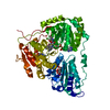
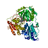
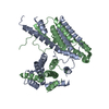
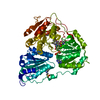
Crystal Structure of Escherichia coli MazG, the Regulator of Nutritional Stress Response
Components
Protein mazG
Keywords
HYDROLASE / tandem repeat domain
Function / homology
Function and homology information
ATP diphosphatase / TTP catabolic process / UTP catabolic process / dTTP catabolic process / dATP catabolic process / nucleoside triphosphate diphosphatase activity / dUTP catabolic process / ATP diphosphatase activity / dGTP catabolic process / cellular response to starvation ...ATP diphosphatase / TTP catabolic process / UTP catabolic process / dTTP catabolic process / dATP catabolic process / nucleoside triphosphate diphosphatase activity / dUTP catabolic process / ATP diphosphatase activity / dGTP catabolic process / cellular response to starvation / ATP binding / metal ion binding Similarity search - Function
Method to determine structure: MOLECULAR REPLACEMENT / Resolution: 3→33.79 Å / Cor.coef. Fo:Fc: 0.914 / Cor.coef. Fo:Fc free: 0.75 / SU B: 22.437 / SU ML: 0.44 / Cross valid method: THROUGHOUT / ESU R Free: 0.604 / Stereochemistry target values: MAXIMUM LIKELIHOOD / Details: HYDROGENS HAVE BEEN ADDED IN THE RIDING POSITIONS
Rfactor
Num. reflection
% reflection
Selection details
Rfree
0.3451
584
4.9 %
RANDOM
Rwork
0.21581
-
-
-
obs
0.22233
11405
93.86 %
-
all
-
12574
-
-
Solvent computation
Ion probe radii: 0.8 Å / Shrinkage radii: 0.8 Å / VDW probe radii: 1.2 Å / Solvent model: MASK
Displacement parameters
Biso mean: 44.569 Å2
Baniso -1
Baniso -2
Baniso -3
1-
0.05 Å2
0 Å2
0 Å2
2-
-
0.06 Å2
0 Å2
3-
-
-
-0.11 Å2
Refinement step
Cycle: LAST / Resolution: 3→33.79 Å
Protein
Nucleic acid
Ligand
Solvent
Total
Num. atoms
3613
0
32
13
3658
Refine LS restraints
Refine-ID
Type
Dev ideal
Dev ideal target
Number
X-RAY DIFFRACTION
r_bond_refined_d
0.041
0.022
3690
X-RAY DIFFRACTION
r_angle_refined_deg
3.742
1.97
4983
X-RAY DIFFRACTION
r_dihedral_angle_1_deg
13.78
5
439
X-RAY DIFFRACTION
r_dihedral_angle_2_deg
38.963
24.537
205
X-RAY DIFFRACTION
r_dihedral_angle_3_deg
26.635
15
668
X-RAY DIFFRACTION
r_dihedral_angle_4_deg
24.193
15
36
X-RAY DIFFRACTION
r_chiral_restr
0.25
0.2
549
X-RAY DIFFRACTION
r_gen_planes_refined
0.013
0.02
2815
X-RAY DIFFRACTION
r_nbd_refined
0.387
0.2
2614
X-RAY DIFFRACTION
r_nbtor_refined
0.378
0.2
2509
X-RAY DIFFRACTION
r_xyhbond_nbd_refined
0.275
0.2
279
X-RAY DIFFRACTION
r_symmetry_vdw_refined
0.366
0.2
54
X-RAY DIFFRACTION
r_symmetry_hbond_refined
0.338
0.2
2
X-RAY DIFFRACTION
r_mcbond_it
1.43
1.5
2309
X-RAY DIFFRACTION
r_mcangle_it
2.351
2
3548
X-RAY DIFFRACTION
r_scbond_it
3.8
3
1590
X-RAY DIFFRACTION
r_scangle_it
5.986
4.5
1435
LS refinement shell
Resolution: 3→3.054 Å / Total num. of bins used: 20
Rfactor
Num. reflection
% reflection
Rfree
0.354
30
-
Rwork
0.226
582
-
obs
-
-
67.11 %
+
About Yorodumi
-
News
-
Feb 9, 2022. New format data for meta-information of EMDB entries
New format data for meta-information of EMDB entries
Version 3 of the EMDB header file is now the official format.
The previous official version 1.9 will be removed from the archive.
In the structure databanks used in Yorodumi, some data are registered as the other names, "COVID-19 virus" and "2019-nCoV". Here are the details of the virus and the list of structure data.
Jan 31, 2019. EMDB accession codes are about to change! (news from PDBe EMDB page)
EMDB accession codes are about to change! (news from PDBe EMDB page)
The allocation of 4 digits for EMDB accession codes will soon come to an end. Whilst these codes will remain in use, new EMDB accession codes will include an additional digit and will expand incrementally as the available range of codes is exhausted. The current 4-digit format prefixed with “EMD-” (i.e. EMD-XXXX) will advance to a 5-digit format (i.e. EMD-XXXXX), and so on. It is currently estimated that the 4-digit codes will be depleted around Spring 2019, at which point the 5-digit format will come into force.
The EM Navigator/Yorodumi systems omit the EMD- prefix.
Related info.:Q: What is EMD? / ID/Accession-code notation in Yorodumi/EM Navigator
Yorodumi is a browser for structure data from EMDB, PDB, SASBDB, etc.
This page is also the successor to EM Navigator detail page, and also detail information page/front-end page for Omokage search.
The word "yorodu" (or yorozu) is an old Japanese word meaning "ten thousand". "mi" (miru) is to see.
Related info.:EMDB / PDB / SASBDB / Comparison of 3 databanks / Yorodumi Search / Aug 31, 2016. New EM Navigator & Yorodumi / Yorodumi Papers / Jmol/JSmol / Function and homology information / Changes in new EM Navigator and Yorodumi
 Movie
Movie Controller

 Yorodumi
 Open data
 Basic information
 Components
 Keywords
 Function and homology information
 X-RAY DIFFRACTION /
  Authors
 Citation
 Structure visualization
 Downloads & links
 Other downloads


 PDBj
PDBj Assembly

 Mass: 24.305 Da / Num. of mol.: 1 / Source method: obtained synthetically / Formula: Mg
 Mass: 507.181 Da / Num. of mol.: 1 / Source method: obtained synthetically / Formula: C10H16N5O13P3 / Comment: ATP, energy-carrying molecule*YM
 Mass: 18.015 Da / Num. of mol.: 13 / Source method: isolated from a natural source / Formula: H2O
 Sample preparation
 / Beamline: 6C1 / Wavelength: 1.23985 Å
 Processing