 Movie
Movie Controller

[English] 日本語
 Yorodumi
- PDB-1yhy: Crystal structure of Arabidopsis thaliana Acetohydroxyacid syntha... -
+ Open data

- Basic information
| Entry | Database: PDB / ID: 1yhy | ||||||
|---|---|---|---|---|---|---|---|
| Title | Crystal structure of Arabidopsis thaliana Acetohydroxyacid synthase In Complex With A Sulfonylurea Herbicide, Metsulfuron methyl | ||||||
 Components Components | Acetolactate synthase | ||||||
 Keywords Keywords | TRANSFERASE / Acetohydroxyacid synthase / acetolactate synthase / herbicide / sulfonylurea / thiamin diphosphate / FAD / inhibitor / cysteine-s-dioxide / CHES / metsulfuron methyl | ||||||
| Function / homology |  Function and homology information Function and homology informationacetolactate synthase / acetolactate synthase complex / acetolactate synthase activity / L-valine biosynthetic process / : / thiamine pyrophosphate binding / response to herbicide / chloroplast stroma / chloroplast / flavin adenine dinucleotide binding / magnesium ion binding Similarity search - Function | ||||||
| Biological species |  | ||||||
| Method |  X-RAY DIFFRACTION / SYNCHROTRON / FOURIER SYNTHESIS / Resolution: 2.7 Å X-RAY DIFFRACTION / SYNCHROTRON / FOURIER SYNTHESIS / Resolution: 2.7 Å | ||||||
 Authors Authors | McCourt, J.A. / Pang, S.S. / King-Scott, J. / Guddat, L.W. / Duggleby, R.G. | ||||||
 Citation Citation | Journal: Proc.Natl.Acad.Sci.Usa / Year: 2006 Title: Herbicide-binding sites revealed in the structure of plant acetohydroxyacid synthase Authors: McCourt, J.A. / Pang, S.S. / King-Scott, J. / Guddat, L.W. / Duggleby, R.G. | ||||||
| History |
|
- Structure visualization
| Structure viewer | Molecule: MolmilJmol/JSmol |
|---|
- Downloads & links
-Download
| PDBx/mmCIF format | 1yhy.cif.gz | 137.5 KB | Display | PDBx/mmCIF format |
|---|---|---|---|---|
| PDB format | pdb1yhy.ent.gz | 101.9 KB | Display | PDB format |
| PDBx/mmJSON format | 1yhy.json.gz | Tree view | PDBx/mmJSON format | |
| Others |  Other downloads Other downloads |
-Validation report
| Arichive directory | https://data.pdbj.org/pub/pdb/validation_reports/yh/1yhyftp://data.pdbj.org/pub/pdb/validation_reports/yh/1yhy | HTTPS FTP |
|---|
-Related structure data
| Related structure data |  1ybhSC  1yhzC  1yi0C  1yi1C  1z8nC C: citing same article ( S: Starting model for refinement |
|---|---|
| Similar structure data |


-Links
 PDBj
PDBj


- Assembly
| Deposited unit | 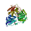
| ||||||||
|---|---|---|---|---|---|---|---|---|---|
| 1 | 
| ||||||||
| 2 | 
| ||||||||
| Unit cell |
| ||||||||
| Details | The biological unit is a tetramer |
-Components
-Protein , 1 types, 1 molecules A
| #1: Protein | Mass: 64591.664 Da / Num. of mol.: 1 / Fragment: residues 86-667 Source method: isolated from a genetically manipulated source Source: (gene. exp.)  |
|---|
-Non-polymers , 6 types, 280 molecules 


| #2: Chemical | ChemComp-MG /  Mass: 24.305 Da / Num. of mol.: 1 / Source method: obtained synthetically / Formula: Mg Mass: 24.305 Da / Num. of mol.: 1 / Source method: obtained synthetically / Formula: Mg |
|---|---|
| #3: Chemical | ChemComp-1MM /  Mass: 381.364 Da / Num. of mol.: 1 / Source method: obtained synthetically / Formula: C14H15N5O6S Mass: 381.364 Da / Num. of mol.: 1 / Source method: obtained synthetically / Formula: C14H15N5O6S |
| #4: Chemical | ChemComp-NHE /  Mass: 207.290 Da / Num. of mol.: 1 / Source method: obtained synthetically / Formula: C8H17NO3S / Comment: pH buffer*YM Mass: 207.290 Da / Num. of mol.: 1 / Source method: obtained synthetically / Formula: C8H17NO3S / Comment: pH buffer*YM |
| #5: Chemical | ChemComp-FAD /  Mass: 785.550 Da / Num. of mol.: 1 / Source method: obtained synthetically / Formula: C27H33N9O15P2 / Comment: FAD*YM Mass: 785.550 Da / Num. of mol.: 1 / Source method: obtained synthetically / Formula: C27H33N9O15P2 / Comment: FAD*YM |
| #6: Chemical | ChemComp-P22 /  Mass: 206.028 Da / Num. of mol.: 1 / Source method: obtained synthetically / Formula: C2H8O7P2 Mass: 206.028 Da / Num. of mol.: 1 / Source method: obtained synthetically / Formula: C2H8O7P2 |
| #7: Water | ChemComp-HOH / Mass: 18.015 Da / Num. of mol.: 275 / Source method: isolated from a natural source / Formula: H2O |
-Details
| Has protein modification | Y |
|---|
-Experimental details
-Experiment
| Experiment | Method: X-RAY DIFFRACTION / Number of used crystals: 1 |
|---|
- Sample preparation
| Crystal | Density Matthews: 6.6 Å3/Da / Density % sol: 80.6 % |
|---|---|
| Crystal grow | Temperature: 290 K / Method: vapor diffusion, hanging drop Details: Tris, FAD, DTT, ThDP, magnesium chloride, metsulfuron methyl,CHES, lithium sufate, potassium sodium tartrate , pH 9.0-9.8, VAPOR DIFFUSION, HANGING DROP, temperature 290K |
-Data collection
| Diffraction | Mean temperature: 100 K |
|---|---|
| Diffraction source | Source: SYNCHROTRON / Site: APS  / Beamline: 14-BM-D / Wavelength: 1 Å / Beamline: 14-BM-D / Wavelength: 1 Å |
| Detector | Type: ADSC QUANTUM 4 / Detector: CCD / Date: Dec 12, 2003 / Details: mirrors |
| Radiation | Monochromator: GE(III) / Protocol: SINGLE WAVELENGTH / Monochromatic (M) / Laue (L): M / Scattering type: x-ray |
| Radiation wavelength | Wavelength: 1 Å / Relative weight: 1 |
| Reflection | Resolution: 2.7→50 Å / Num. all: 45178 / Num. obs: 45178 / % possible obs: 93.8 % / Observed criterion σ(F): 0 / Observed criterion σ(I): -3 / Redundancy: 3.9 % / Rmerge(I) obs: 0.099 / Rsym value: 0.099 / Net I/σ(I): 7.3 |
| Reflection shell | Resolution: 2.7→2.8 Å / Redundancy: 4.3 % / Mean I/σ(I) obs: 3.1 / Num. unique all: 4506 / % possible all: 95.5 |
- Processing
| Software |
| |||||||||||||||||||||||||
|---|---|---|---|---|---|---|---|---|---|---|---|---|---|---|---|---|---|---|---|---|---|---|---|---|---|---|
| Refinement | Method to determine structure: FOURIER SYNTHESIS Starting model: 1YBH Resolution: 2.7→50 Å / Isotropic thermal model: Isotropic / Cross valid method: THROUGHOUT / σ(F): 0 / Stereochemistry target values: Engh & Huber
| |||||||||||||||||||||||||
| Displacement parameters | Biso mean: 45.362 Å2 | |||||||||||||||||||||||||
| Refine analyze |
| |||||||||||||||||||||||||
| Refinement step | Cycle: LAST / Resolution: 2.7→50 Å
| |||||||||||||||||||||||||
| LS refinement shell | Resolution: 2.7→2.8 Å
|
