 Movie
Movie Controller


+ Open data

- Basic information
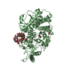
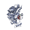
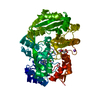
| Entry | Database: PDB / ID: 3ck7 | |||||||||
|---|---|---|---|---|---|---|---|---|---|---|
| Title | B. thetaiotaomicron SusD with alpha-cyclodextrin | |||||||||
 Components Components | SusD | |||||||||
 Keywords Keywords | SUGAR BINDING PROTEIN / TPR repeat / carbohydrate binding / starch binding | |||||||||
| Function / homology |  Function and homology information Function and homology informationstarch metabolic process / starch catabolic process / starch binding / outer membrane / cell outer membrane / calcium ion binding / identical protein binding Similarity search - Function | |||||||||
| Biological species |  Bacteroides thetaiotaomicron (bacteria) Bacteroides thetaiotaomicron (bacteria) | |||||||||
| Method |  X-RAY DIFFRACTION / MOLECULAR REPLACEMENT / molecular replacement / Resolution: 2.1 Å X-RAY DIFFRACTION / MOLECULAR REPLACEMENT / molecular replacement / Resolution: 2.1 Å | |||||||||
 Authors Authors | Koropatkin, N.M. / Martens, E.C. / Gordon, J.I. / Smith, T.J. | |||||||||
 Citation Citation | Journal: Structure / Year: 2008 Title: Starch catabolism by a prominent human gut symbiont is directed by the recognition of amylose helices. Authors: Koropatkin, N.M. / Martens, E.C. / Gordon, J.I. / Smith, T.J. | |||||||||
| History |
|
- Structure visualization
| Structure viewer | Molecule: MolmilJmol/JSmol |
|---|
- Downloads & links
-Download
| PDBx/mmCIF format | 3ck7.cif.gz | 431.2 KB | Display | PDBx/mmCIF format |
|---|---|---|---|---|
| PDB format | pdb3ck7.ent.gz | 348.1 KB | Display | PDB format |
| PDBx/mmJSON format | 3ck7.json.gz | Tree view | PDBx/mmJSON format | |
| Others |  Other downloads Other downloads |
-Validation report
| Arichive directory | https://data.pdbj.org/pub/pdb/validation_reports/ck/3ck7ftp://data.pdbj.org/pub/pdb/validation_reports/ck/3ck7 | HTTPS FTP |
|---|
-Related structure data
| Related structure data |  3ck8SC  3ck9C  3ckbC  3ckcC S: Starting model for refinement C: citing same article ( |
|---|---|
| Similar structure data |


-Links
 PDBj
PDBj


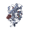
- Assembly
| Deposited unit | 
| ||||||||
|---|---|---|---|---|---|---|---|---|---|
| 1 | 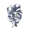
| ||||||||
| 2 | 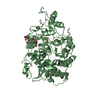
| ||||||||
| 3 | 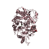
| ||||||||
| 4 | 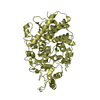
| ||||||||
| Unit cell |
|
-Components
| #1: Protein | Mass: 59783.340 Da / Num. of mol.: 4 / Fragment: UNP residues 26-551 Source method: isolated from a genetically manipulated source Source: (gene. exp.) Bacteroides thetaiotaomicron (bacteria)Strain: VPI-5482 / Gene: SusD / Plasmid: pET-28a / Production host: #2: Polysaccharide | 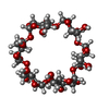  Source method: isolated from a genetically manipulated source Details: cyclic oligosaccharide / References: alpha-cyclodextrin #3: Chemical | ChemComp-CA /   Mass: 40.078 Da / Num. of mol.: 4 / Source method: obtained synthetically / Formula: Ca Mass: 40.078 Da / Num. of mol.: 4 / Source method: obtained synthetically / Formula: Ca#4: Water | ChemComp-HOH / |  Mass: 18.015 Da / Num. of mol.: 1332 / Source method: isolated from a natural source / Formula: H2O Mass: 18.015 Da / Num. of mol.: 1332 / Source method: isolated from a natural source / Formula: H2OSequence details | THE ORIGINAL SUSD GENE SEQUENCE DEPOSITED BY WASHINGTON UNIVERSITY (FROM JEFFREY I GORDON'S ...THE ORIGINAL SUSD GENE SEQUENCE DEPOSITED BY WASHINGTON | |
|---|
-Experimental details
-Experiment
| Experiment | Method: X-RAY DIFFRACTION / Number of used crystals: 1 |
|---|
- Sample preparation
| Crystal | Density Matthews: 2.56 Å3/Da / Density % sol: 51.95 % |
|---|---|
| Crystal grow | Temperature: 298 K / Method: seeding in batch / pH: 8.5 Details: 50mM Tris pH 8.5, 100mM sodium acetate, 15% PEG 4000, 2.5mM alpha-cyclodextrin, seeding in batch, temperature 298K |
-Data collection
| Diffraction | Mean temperature: 150 K |
|---|---|
| Diffraction source | Source: ROTATING ANODE / Type: OTHER / Wavelength: 1.5418 |
| Detector | Type: BRUKER SMART 6000 / Detector: CCD / Date: Oct 1, 2007 / Details: mirrors |
| Radiation | Monochromator: mirrors / Protocol: SINGLE WAVELENGTH / Monochromatic (M) / Laue (L): M / Scattering type: x-ray |
| Radiation wavelength | Wavelength: 1.5418 Å / Relative weight: 1 |
| Reflection | Resolution: 2.1→67.5 Å / Num. all: 127524 / Num. obs: 127524 / % possible obs: 93.4 % / Observed criterion σ(F): 0 / Observed criterion σ(I): 0 / Redundancy: 4.5 % / Rsym value: 0.096 / Net I/σ(I): 6.9 |
| Reflection shell | Resolution: 2.1→2.21 Å / Redundancy: 1.5 % / Mean I/σ(I) obs: 1.9 / Num. unique all: 16027 / Rsym value: 0.213 / % possible all: 81.2 |
-Phasing
| Phasing | Method: molecular replacement |
|---|
- Processing
| Software |
| ||||||||||||||||||||||||||||
|---|---|---|---|---|---|---|---|---|---|---|---|---|---|---|---|---|---|---|---|---|---|---|---|---|---|---|---|---|---|
| Refinement | Method to determine structure: MOLECULAR REPLACEMENT Starting model: PDB entry 3CK8 Resolution: 2.1→67.5 Å / FOM work R set: 0.865 / Cross valid method: THROUGHOUT / σ(F): 0 / σ(I): 0 / Stereochemistry target values: Engh & Huber
| ||||||||||||||||||||||||||||
| Solvent computation | Bsol: 47.812 Å2 | ||||||||||||||||||||||||||||
| Displacement parameters | Biso mean: 12.357 Å2
| ||||||||||||||||||||||||||||
| Refinement step | Cycle: LAST / Resolution: 2.1→67.5 Å
| ||||||||||||||||||||||||||||
| Refine LS restraints |
| ||||||||||||||||||||||||||||
| Xplor file |
|
