 Movie
Movie Controller

[English] 日本語
 Yorodumi
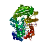
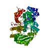
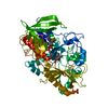
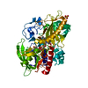
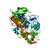
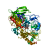
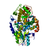
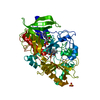
- PDB-3b6d: Crystal Structure of Streptomyces Cholesterol Oxidase H447Q/E361Q... -
+ Open data

- Basic information
| Entry | Database: PDB / ID: 3b6d | ||||||
|---|---|---|---|---|---|---|---|
| Title | Crystal Structure of Streptomyces Cholesterol Oxidase H447Q/E361Q mutant (1.2A) | ||||||
 Components Components | Cholesterol oxidase | ||||||
 Keywords Keywords | OXIDOREDUCTASE / FLAVOENZYME / FLAVIN / FLAVIN ACTIVATION / CHOLESTEROL OXIDASE / Cholesterol metabolism / FAD / Flavoprotein / Lipid metabolism / Secreted / Steroid metabolism | ||||||
| Function / homology |  Function and homology information Function and homology informationcholesterol oxidase / cholesterol oxidase activity / steroid Delta-isomerase / steroid Delta-isomerase activity / cholesterol catabolic process / flavin adenine dinucleotide binding / extracellular region Similarity search - Function | ||||||
| Biological species |  Streptomyces sp. (bacteria) Streptomyces sp. (bacteria) | ||||||
| Method |  X-RAY DIFFRACTION / FOURIER SYNTHESIS / Resolution: 1.2 Å X-RAY DIFFRACTION / FOURIER SYNTHESIS / Resolution: 1.2 Å | ||||||
 Authors Authors | Lyubimov, A.Y. / Vrielink, A. | ||||||
 Citation Citation | Journal: Protein Sci. / Year: 2007 Title: Distortion of flavin geometry is linked to ligand binding in cholesterol oxidase Authors: Lyubimov, A.Y. / Heard, K. / Tang, H. / Sampson, N.S. / Vrielink, A. | ||||||
| History |
|
- Structure visualization
| Structure viewer | Molecule: MolmilJmol/JSmol |
|---|
- Downloads & links
-Download
| PDBx/mmCIF format | 3b6d.cif.gz | 340.4 KB | Display | PDBx/mmCIF format |
|---|---|---|---|---|
| PDB format | pdb3b6d.ent.gz | 278.8 KB | Display | PDB format |
| PDBx/mmJSON format | 3b6d.json.gz | Tree view | PDBx/mmJSON format | |
| Others |  Other downloads Other downloads |
-Validation report
| Arichive directory | https://data.pdbj.org/pub/pdb/validation_reports/b6/3b6dftp://data.pdbj.org/pub/pdb/validation_reports/b6/3b6d | HTTPS FTP |
|---|
-Related structure data
| Related structure data | 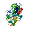 3b3rC  1mxtS C: citing same article ( S: Starting model for refinement |
|---|---|
| Similar structure data |


-Links
 PDBj
PDBj


- Assembly
| Deposited unit | 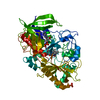
| ||||||||
|---|---|---|---|---|---|---|---|---|---|
| 1 |
| ||||||||
| Unit cell |
|
-Components
| #1: Protein | Mass: 54958.676 Da / Num. of mol.: 1 / Mutation: H447Q/E361Q Source method: isolated from a genetically manipulated source Details: FAD COFACTOR NON-COVALENTLY BOUND TO THE ENZYME / Source: (gene. exp.) Streptomyces sp. (bacteria) / Strain: SA-COO / Gene: choA / Plasmid: PCO202 / Production host: |
|---|---|
| #2: Chemical | ChemComp-SO4 /   Mass: 96.063 Da / Num. of mol.: 1 / Source method: obtained synthetically / Formula: SO4 Mass: 96.063 Da / Num. of mol.: 1 / Source method: obtained synthetically / Formula: SO4 |
| #3: Chemical | ChemComp-FAE / 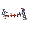  Mass: 786.558 Da / Num. of mol.: 1 / Source method: obtained synthetically / Formula: C27H34N9O15P2 Mass: 786.558 Da / Num. of mol.: 1 / Source method: obtained synthetically / Formula: C27H34N9O15P2 |
| #4: Water | ChemComp-HOH /  Mass: 18.015 Da / Num. of mol.: 560 / Source method: isolated from a natural source / Formula: H2O Mass: 18.015 Da / Num. of mol.: 560 / Source method: isolated from a natural source / Formula: H2O |
-Experimental details
-Experiment
| Experiment | Method: X-RAY DIFFRACTION / Number of used crystals: 1 |
|---|
- Sample preparation
| Crystal | Density Matthews: 2.08 Å3/Da / Density % sol: 40.9 % |
|---|---|
| Crystal grow | Temperature: 291 K / Method: vapor diffusion, hanging drop / pH: 5.2 Details: 10% PEG 8000, 75mM MnSO4, 100mM CACODYLATE pH 5.2, VAPOR DIFFUSION, HANGING DROP, temperature 291K |
-Data collection
| Diffraction | Mean temperature: 100 K |
|---|---|
| Diffraction source | Source: ROTATING ANODE / Type: RIGAKU MICROMAX-007 HF / Wavelength: 1.5 Å |
| Detector | Type: RIGAKU RAXIS IV++ / Detector: IMAGE PLATE |
| Radiation | Protocol: SINGLE WAVELENGTH / Monochromatic (M) / Laue (L): M / Scattering type: x-ray |
| Radiation wavelength | Wavelength: 1.5 Å / Relative weight: 1 |
| Reflection | Resolution: 1.2→60.97 Å / Num. obs: 132078 / % possible obs: 94.1 % / Redundancy: 3.3 % / Rmerge(I) obs: 0.1 / Net I/σ(I): 11.9 |
| Reflection shell | Resolution: 1.2→1.22 Å / Redundancy: 1.3 % / Rmerge(I) obs: 0.196 / Mean I/σ(I) obs: 3.2 / % possible all: 74.5 |
- Processing
| Software |
| |||||||||||||||||||||||||||||||||
|---|---|---|---|---|---|---|---|---|---|---|---|---|---|---|---|---|---|---|---|---|---|---|---|---|---|---|---|---|---|---|---|---|---|---|
| Refinement | Method to determine structure: FOURIER SYNTHESIS Starting model: PDB ENTRY 1MXT; ADP, WATERS, LIGANDS AND ACTIVE SITE SIDECHAINS REMOVED FROM STARTING MODEL Resolution: 1.2→38.03 Å / Num. parameters: 43986 / Num. restraintsaints: 62007 / Cross valid method: FREE R / σ(F): 0 / Stereochemistry target values: Engh & Huber
| |||||||||||||||||||||||||||||||||
| Refine analyze | Num. disordered residues: 73 / Occupancy sum hydrogen: 3714 / Occupancy sum non hydrogen: 4427.84 | |||||||||||||||||||||||||||||||||
| Refinement step | Cycle: LAST / Resolution: 1.2→38.03 Å
| |||||||||||||||||||||||||||||||||
| Refine LS restraints |
|
