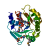
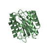
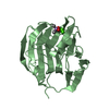
- PDB-3cjl: Crystal structure of a protein of unknown function (eca1910) from... -
+
Open data
ID or keywords:
Loading...
-
Basic information
Entry
Database: PDB / ID: 3cjl
Title
Crystal structure of a protein of unknown function (eca1910) from pectobacterium atrosepticum scri1043 at 2.20 A resolution
Components
Domain of unknown function
Keywords
UNKNOWN FUNCTION / Structural genomics / Joint Center for Structural Genomics / JCSG / Protein Structure Initiative / PSI-2
Function / homology
Protein of unknown function DUF3861 / Protein of unknown function DUF3861 / DUF3861 domain superfamily / Domain of Unknown Function with PDB structure (DUF3861) / Ubiquitin-like (UB roll) / Roll / Alpha Beta / Uncharacterized protein
Function and homology information
Biological species
Pectobacterium atrosepticum SCRI1043 (bacteria)
Method
X-RAY DIFFRACTION / SYNCHROTRON / SAD / Resolution: 2.2 Å
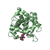
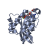
Mass: 18.015 Da / Num. of mol.: 56 / Source method: isolated from a natural source / Formula: H2O
Sequence details
THE CONSTRUCT WAS EXPRESSED WITH A PURIFICATION TAG MGSDKIHHHHHHENLYFQG. THE TAG WAS REMOVED WITH ...THE CONSTRUCT WAS EXPRESSED WITH A PURIFICATION TAG MGSDKIHHHHHHENLYFQG. THE TAG WAS REMOVED WITH TEV PROTEASE LEAVING ONLY A GLYCINE (0) FOLLOWED BY THE TARGET SEQUENCE.
-
Experimental details
-
Experiment
Experiment
Method: X-RAY DIFFRACTION / Number of used crystals: 1
-
Sample preparation
Crystal
Density Matthews: 3.19 Å3/Da / Density % sol: 61.46 %
Crystal grow
Temperature: 277 K / Method: vapor diffusion, sitting drop / pH: 7.5 Details: NANODROP, 1.4M Na3Citrate, 0.1M HEPES pH 7.5, VAPOR DIFFUSION, SITTING DROP, temperature 277K
Monochromator: Double crystal / Protocol: SINGLE WAVELENGTH / Monochromatic (M) / Laue (L): M / Scattering type: x-ray
Radiation wavelength
Wavelength: 0.97908 Å / Relative weight: 1
Reflection
Resolution: 2.2→29.123 Å / Num. obs: 14004 / % possible obs: 99.9 % / Redundancy: 7.3 % / Biso Wilson estimate: 39.316 Å2 / Rmerge(I) obs: 0.106 / Rsym value: 0.106 / Net I/σ(I): 5.1
Reflection shell
Diffraction-ID: 1
Resolution (Å)
Redundancy (%)
Rmerge(I) obs
Mean I/σ(I) obs
Num. measured all
Num. unique all
Rsym value
% possible all
2.2-2.26
7.4
0.716
1.1
7582
1022
0.716
100
2.26-2.32
7.5
0.611
1.3
7596
1017
0.611
100
2.32-2.39
7.5
0.487
1.6
7068
948
0.487
100
2.39-2.46
7.4
0.416
1.9
7240
973
0.416
100
2.46-2.54
7.5
0.348
2.2
6727
899
0.348
100
2.54-2.63
7.4
0.282
2.7
6562
882
0.282
100
2.63-2.73
7.5
0.229
3.4
6523
874
0.229
100
2.73-2.84
7.5
0.183
4.2
6038
809
0.183
100
2.84-2.97
7.5
0.164
4.6
5954
799
0.164
100
2.97-3.11
7.5
0.129
5.7
5755
768
0.129
100
3.11-3.28
7.4
0.111
6.4
5398
727
0.111
100
3.28-3.48
7.4
0.092
7.4
5047
683
0.092
100
3.48-3.72
7.4
0.082
8.1
4727
642
0.082
100
3.72-4.02
7.3
0.073
9.1
4350
593
0.073
100
4.02-4.4
7.2
0.07
9.8
4060
563
0.07
100
4.4-4.92
7.2
0.068
9.4
3555
497
0.068
100
4.92-5.68
6.8
0.079
8.1
3101
457
0.079
100
5.68-6.96
6.3
0.09
7.2
2431
383
0.09
100
6.96-9.84
6.1
0.065
9.2
1837
302
0.065
100
9.84-29.123
5.6
0.065
9.1
930
166
0.065
94.8
-
Phasing
Phasing
Method: SAD
-
Processing
Software
Name
Version
Classification
NB
REFMAC
5.2.0019
refinement
PHENIX
refinement
SOLVE
phasing
MolProbity
3beta29
modelbuilding
SCALA
datascaling
PDB_EXTRACT
3
dataextraction
MAR345
CCD
datacollection
MOSFLM
datareduction
Refinement
Method to determine structure: SAD / Resolution: 2.2→29.123 Å / Cor.coef. Fo:Fc: 0.954 / Cor.coef. Fo:Fc free: 0.938 / SU B: 9.998 / SU ML: 0.125 / TLS residual ADP flag: LIKELY RESIDUAL / Cross valid method: THROUGHOUT / σ(F): 0 / ESU R: 0.194 / ESU R Free: 0.164 Stereochemistry target values: MAXIMUM LIKELIHOOD WITH PHASES Details: 1. HYDROGENS HAVE BEEN ADDED IN THE RIDING POSITIONS. 2. A MET-INHIBITION PROTOCOL WAS USED FOR SELENOMETHIONINE INCORPORATION DURING PROTEIN EXPRESSION. THE OCCUPANCY OF THE SE ATOMS IN THE ...Details: 1. HYDROGENS HAVE BEEN ADDED IN THE RIDING POSITIONS. 2. A MET-INHIBITION PROTOCOL WAS USED FOR SELENOMETHIONINE INCORPORATION DURING PROTEIN EXPRESSION. THE OCCUPANCY OF THE SE ATOMS IN THE MSE RESIDUES WAS REDUCED TO 0.75 FOR THE REDUCED SCATTERING POWER DUE TO PARTIAL S-MET INCORPORATION. 3. ATOM RECORD CONTAINS RESIDUAL B FACTORS ONLY.
Rfactor
Num. reflection
% reflection
Selection details
Rfree
0.22
695
5 %
RANDOM
Rwork
0.194
-
-
-
obs
0.195
13985
99.94 %
-
Solvent computation
Ion probe radii: 0.8 Å / Shrinkage radii: 0.8 Å / VDW probe radii: 1.2 Å / Solvent model: MASK
In the structure databanks used in Yorodumi, some data are registered as the other names, "COVID-19 virus" and "2019-nCoV". Here are the details of the virus and the list of structure data.
Jan 31, 2019. EMDB accession codes are about to change! (news from PDBe EMDB page)
EMDB accession codes are about to change! (news from PDBe EMDB page)
The allocation of 4 digits for EMDB accession codes will soon come to an end. Whilst these codes will remain in use, new EMDB accession codes will include an additional digit and will expand incrementally as the available range of codes is exhausted. The current 4-digit format prefixed with “EMD-” (i.e. EMD-XXXX) will advance to a 5-digit format (i.e. EMD-XXXXX), and so on. It is currently estimated that the 4-digit codes will be depleted around Spring 2019, at which point the 5-digit format will come into force.
The EM Navigator/Yorodumi systems omit the EMD- prefix.
Related info.:Q: What is EMD? / ID/Accession-code notation in Yorodumi/EM Navigator
Yorodumi is a browser for structure data from EMDB, PDB, SASBDB, etc.
This page is also the successor to EM Navigator detail page, and also detail information page/front-end page for Omokage search.
The word "yorodu" (or yorozu) is an old Japanese word meaning "ten thousand". "mi" (miru) is to see.
Related info.:EMDB / PDB / SASBDB / Comparison of 3 databanks / Yorodumi Search / Aug 31, 2016. New EM Navigator & Yorodumi / Yorodumi Papers / Jmol/JSmol / Function and homology information / Changes in new EM Navigator and Yorodumi
 Movie
Movie Controller

 Yorodumi
 Open data
 Basic information
 Components
 Keywords
 Function and homology information
 Pectobacterium atrosepticum SCRI1043 (bacteria)
 X-RAY DIFFRACTION /
  Authors
 Citation
 Structure visualization
 Downloads & links
 Other downloads
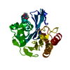


 PDBj
PDBj Assembly

 Mass: 62.068 Da / Num. of mol.: 1 / Source method: obtained synthetically / Formula: C2H6O2
 Mass: 18.015 Da / Num. of mol.: 56 / Source method: isolated from a natural source / Formula: H2O
 Sample preparation
 / Beamline: BL9-2 / Wavelength: 0.97908 Å
 Processing