Mass: 18.015 Da / Num. of mol.: 141 / Source method: isolated from a natural source / Formula: H2O
Has protein modification
N
-
Experimental details
-
Experiment
Experiment
Method: X-RAY DIFFRACTION / Number of used crystals: 1
-
Sample preparation
Crystal
Density Matthews: 2.87 Å3/Da / Density % sol: 57.18 %
Crystal grow
Method: vapor diffusion, hanging drop / pH: 6.5 Details: VAPOR DIFFUSION, HANGING DROP, 7 mg/ml in 20 mM BES, 1 mM EDTA, 0.5 mM DTT, pH 6.5, 50 mM NBG, 2.5 mM AVF (AVE2865)
-
Data collection
Diffraction
Mean temperature: 100 K
Diffraction source
Source: SYNCHROTRON / Site: SLS / Beamline: X06SA
Radiation
Protocol: SINGLE WAVELENGTH / Monochromatic (M) / Laue (L): M / Scattering type: x-ray
Radiation wavelength
Relative weight: 1
Reflection
Resolution: 3.3→20 Å / Num. obs: 25212 / % possible obs: 80.8 %
-
Processing
Software
Name
Version
Classification
NB
REFMAC
5.2.0019
refinement
PDB_EXTRACT
3.004
dataextraction
XDS
datareduction
XSCALE
datascaling
Refinement
Resolution: 3.3→19.47 Å / Cor.coef. Fo:Fc: 0.925 / Cor.coef. Fo:Fc free: 0.807 / SU B: 28.423 / SU ML: 0.477 / Cross valid method: THROUGHOUT / σ(F): 0 / ESU R Free: 0.72 / Stereochemistry target values: MAXIMUM LIKELIHOOD
Rfactor
Num. reflection
% reflection
Selection details
Rfree
0.271
1257
5 %
RANDOM
Rwork
0.176
-
-
-
obs
0.181
25212
81.21 %
-
Solvent computation
Ion probe radii: 0.8 Å / Shrinkage radii: 0.8 Å / VDW probe radii: 1.2 Å / Solvent model: BABINET MODEL WITH MASK
Displacement parameters
Biso mean: 56.085 Å2
Baniso -1
Baniso -2
Baniso -3
1-
0.04 Å2
0.02 Å2
0 Å2
2-
-
0.04 Å2
0 Å2
3-
-
-
-0.06 Å2
Refinement step
Cycle: LAST / Resolution: 3.3→19.47 Å
Protein
Nucleic acid
Ligand
Solvent
Total
Num. atoms
12830
0
124
141
13095
Refine LS restraints
Refine-ID
Type
Dev ideal
Dev ideal target
Number
X-RAY DIFFRACTION
r_bond_refined_d
0.009
0.022
13250
X-RAY DIFFRACTION
r_angle_refined_deg
1.259
1.969
17932
X-RAY DIFFRACTION
r_dihedral_angle_1_deg
6.005
5
1574
X-RAY DIFFRACTION
r_dihedral_angle_2_deg
38.435
24.295
638
X-RAY DIFFRACTION
r_dihedral_angle_3_deg
18.882
15
2360
X-RAY DIFFRACTION
r_dihedral_angle_4_deg
20.061
15
78
X-RAY DIFFRACTION
r_chiral_restr
0.081
0.2
1956
X-RAY DIFFRACTION
r_gen_planes_refined
0.004
0.02
10010
X-RAY DIFFRACTION
r_nbd_refined
0.24
0.2
8031
X-RAY DIFFRACTION
r_nbtor_refined
0.318
0.2
9013
X-RAY DIFFRACTION
r_xyhbond_nbd_refined
0.185
0.2
656
X-RAY DIFFRACTION
r_symmetry_vdw_refined
0.248
0.2
73
X-RAY DIFFRACTION
r_symmetry_hbond_refined
0.152
0.2
6
X-RAY DIFFRACTION
r_mcbond_it
0.354
1.5
8062
X-RAY DIFFRACTION
r_mcangle_it
0.644
2
12744
X-RAY DIFFRACTION
r_scbond_it
0.728
3
5891
X-RAY DIFFRACTION
r_scangle_it
1.22
4.5
5188
LS refinement shell
Resolution: 3.3→3.383 Å / Total num. of bins used: 20
Rfactor
Num. reflection
% reflection
Rfree
0.354
91
-
Rwork
0.227
1794
-
all
-
1885
-
obs
-
-
82.78 %
+
About Yorodumi
-
News
-
Feb 9, 2022. New format data for meta-information of EMDB entries
New format data for meta-information of EMDB entries
Version 3 of the EMDB header file is now the official format.
The previous official version 1.9 will be removed from the archive.
In the structure databanks used in Yorodumi, some data are registered as the other names, "COVID-19 virus" and "2019-nCoV". Here are the details of the virus and the list of structure data.
Jan 31, 2019. EMDB accession codes are about to change! (news from PDBe EMDB page)
EMDB accession codes are about to change! (news from PDBe EMDB page)
The allocation of 4 digits for EMDB accession codes will soon come to an end. Whilst these codes will remain in use, new EMDB accession codes will include an additional digit and will expand incrementally as the available range of codes is exhausted. The current 4-digit format prefixed with “EMD-” (i.e. EMD-XXXX) will advance to a 5-digit format (i.e. EMD-XXXXX), and so on. It is currently estimated that the 4-digit codes will be depleted around Spring 2019, at which point the 5-digit format will come into force.
The EM Navigator/Yorodumi systems omit the EMD- prefix.
Related info.:Q: What is EMD? / ID/Accession-code notation in Yorodumi/EM Navigator
Yorodumi is a browser for structure data from EMDB, PDB, SASBDB, etc.
This page is also the successor to EM Navigator detail page, and also detail information page/front-end page for Omokage search.
The word "yorodu" (or yorozu) is an old Japanese word meaning "ten thousand". "mi" (miru) is to see.
Related info.:EMDB / PDB / SASBDB / Comparison of 3 databanks / Yorodumi Search / Aug 31, 2016. New EM Navigator & Yorodumi / Yorodumi Papers / Jmol/JSmol / Function and homology information / Changes in new EM Navigator and Yorodumi
 Movie
Movie Controller

 Yorodumi
 Open data
 Basic information
 Components
 Keywords
 Function and homology information
 Homo sapiens (human)
 X-RAY DIFFRACTION /
  Authors
 Citation
 Structure visualization
 Downloads & links
 Other downloads
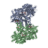


 PDBj
PDBj


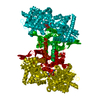
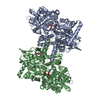
 Assembly
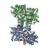

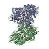
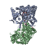
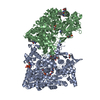
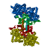
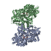
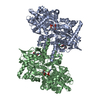
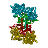
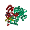
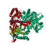
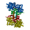
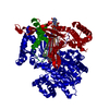
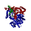
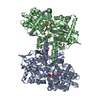
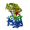
 Spodoptera frugiperda (fall armyworm) / References: UniProt: P06737, glycogen phosphorylase
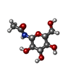
 Type: D-saccharide / Mass: 221.208 Da / Num. of mol.: 2
 Mass: 247.142 Da / Num. of mol.: 2 / Source method: obtained synthetically / Formula: C8H10NO6P
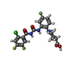
 Mass: 455.815 Da / Num. of mol.: 2 / Source method: obtained synthetically / Formula: C20H17ClF3N3O4
 Mass: 18.015 Da / Num. of mol.: 141 / Source method: isolated from a natural source / Formula: H2O
 Sample preparation
 / Beamline: X06SA
 Processing