 Movie
Movie Controller

[English] 日本語
 Yorodumi
- PDB-1exv: HUMAN LIVER GLYCOGEN PHOSPHORYLASE A COMPLEXED WITH GLCNAC AND CP... -
+ Open data

- Basic information
| Entry | Database: PDB / ID: 1exv | ||||||
|---|---|---|---|---|---|---|---|
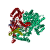
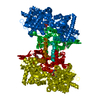
| Title | HUMAN LIVER GLYCOGEN PHOSPHORYLASE A COMPLEXED WITH GLCNAC AND CP-403,700 | ||||||
 Components Components | LIVER GLYCOGEN PHOSPHORYLASE | ||||||
 Keywords Keywords | TRANSFERASE / allosteric site / allosteric binding | ||||||
| Function / homology |  Function and homology information Function and homology informationpurine nucleobase binding / vitamin binding / D-glucose binding / bile acid binding / glycogen phosphorylase / glycogen phosphorylase activity / glycogen catabolic process / glycogen metabolic process / Glycogen breakdown (glycogenolysis) / AMP binding ...purine nucleobase binding / vitamin binding / D-glucose binding / bile acid binding / glycogen phosphorylase / glycogen phosphorylase activity / glycogen catabolic process / glycogen metabolic process / Glycogen breakdown (glycogenolysis) / AMP binding / necroptotic process / response to bacterium / pyridoxal phosphate binding / glucose homeostasis / secretory granule lumen / ficolin-1-rich granule lumen / Neutrophil degranulation / extracellular exosome / extracellular region / ATP binding / identical protein binding / cytoplasm / cytosol Similarity search - Function | ||||||
| Biological species |  Homo sapiens (human) Homo sapiens (human) | ||||||
| Method |  X-RAY DIFFRACTION / Resolution: 2.4 Å X-RAY DIFFRACTION / Resolution: 2.4 Å | ||||||
 Authors Authors | Rath, V.L. / Ammirati, M. / Danley, D.E. / Ekstrom, J.L. / Hynes, T.R. / Olson, T.V. / Hoover, D.J. | ||||||
 Citation Citation | Journal: Chem.Biol. / Year: 2000 Title: Human liver glycogen phosphorylase inhibitors bind at a new allosteric site. Authors: Rath, V.L. / Ammirati, M. / Danley, D.E. / Ekstrom, J.L. / Gibbs, E.M. / Hynes, T.R. / Mathiowetz, A.M. / McPherson, R.K. / Olson, T.V. / Treadway, J.L. / Hoover, D.J. | ||||||
| History |
|
- Structure visualization
| Structure viewer | Molecule: MolmilJmol/JSmol |
|---|
- Downloads & links
-Download
| PDBx/mmCIF format | 1exv.cif.gz | 336.4 KB | Display | PDBx/mmCIF format |
|---|---|---|---|---|
| PDB format | pdb1exv.ent.gz | 266.9 KB | Display | PDB format |
| PDBx/mmJSON format | 1exv.json.gz | Tree view | PDBx/mmJSON format | |
| Others |  Other downloads Other downloads |
-Validation report
| Arichive directory | https://data.pdbj.org/pub/pdb/validation_reports/ex/1exvftp://data.pdbj.org/pub/pdb/validation_reports/ex/1exv | HTTPS FTP |
|---|
-Related structure data


-Links
 PDBj
PDBj


- Assembly
| Deposited unit | 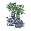
| ||||||||
|---|---|---|---|---|---|---|---|---|---|
| 1 |
| ||||||||
| Unit cell |
| ||||||||
| Details | The biological assembly is a homodimer consisting of 2 identical chains (A and B). |
-Components
-Protein / Sugars , 2 types, 4 molecules AB
| #1: Protein | Mass: 97276.469 Da / Num. of mol.: 2 Source method: isolated from a genetically manipulated source Source: (gene. exp.) Homo sapiens (human) / Organ: LIVER / Plasmid: PBLUEBACII / Cell line (production host): SF9 / Production host:   Spodoptera frugiperda (fall armyworm) / References: UniProt: P06737, glycogen phosphorylase Spodoptera frugiperda (fall armyworm) / References: UniProt: P06737, glycogen phosphorylase#2: Sugar |  Type: D-saccharide / Mass: 221.208 Da / Num. of mol.: 2 Type: D-saccharide / Mass: 221.208 Da / Num. of mol.: 2Source method: isolated from a genetically manipulated source Formula: C8H15NO6 |
|---|
-Non-polymers , 4 types, 339 molecules 


| #3: Chemical |  Mass: 247.142 Da / Num. of mol.: 2 / Source method: obtained synthetically / Formula: C8H10NO6P Mass: 247.142 Da / Num. of mol.: 2 / Source method: obtained synthetically / Formula: C8H10NO6P#4: Chemical |  Mass: 425.865 Da / Num. of mol.: 2 / Source method: obtained synthetically / Formula: C22H20ClN3O4 Mass: 425.865 Da / Num. of mol.: 2 / Source method: obtained synthetically / Formula: C22H20ClN3O4#5: Chemical |  Mass: 118.174 Da / Num. of mol.: 2 / Source method: obtained synthetically / Formula: C6H14O2 / Comment: precipitant*YM Mass: 118.174 Da / Num. of mol.: 2 / Source method: obtained synthetically / Formula: C6H14O2 / Comment: precipitant*YM#6: Water | ChemComp-HOH / | Mass: 18.015 Da / Num. of mol.: 333 / Source method: isolated from a natural source / Formula: H2O |
|---|
-Details
| Has protein modification | N |
|---|
-Experimental details
-Experiment
| Experiment | Method: X-RAY DIFFRACTION / Number of used crystals: 1 |
|---|
- Sample preparation
| Crystal | Density Matthews: 2.86 Å3/Da / Density % sol: 56.97 % |
|---|---|
| Crystal grow | Temperature: 290 K / Method: vapor diffusion, hanging drop / pH: 6 Details: MES, MPD, pH 6.0, VAPOR DIFFUSION, HANGING DROP, temperature 290K |
| Crystal grow | *PLUS Method: unknown |
-Data collection
| Diffraction | Mean temperature: 100 K |
|---|---|
| Diffraction source | Source: ROTATING ANODE / Type: OTHER / Wavelength: 1.5418 |
| Detector | Type: MARRESEARCH / Detector: IMAGE PLATE / Date: Oct 21, 1996 |
| Radiation | Protocol: SINGLE WAVELENGTH / Monochromatic (M) / Laue (L): M / Scattering type: x-ray |
| Radiation wavelength | Wavelength: 1.5418 Å / Relative weight: 1 |
| Reflection | Resolution: 2.4→99 Å / Num. all: 84103 / Num. obs: 84018 / % possible obs: 99.8 % / Observed criterion σ(F): 0 / Observed criterion σ(I): -2 / Redundancy: 4.68 % / Biso Wilson estimate: 31.2 Å2 / Rmerge(I) obs: 0.071 / Net I/σ(I): 19.11 |
| Reflection shell | Resolution: 2.4→2.49 Å / Redundancy: 3 % / Rmerge(I) obs: 0.378 / Num. unique all: 8330 / % possible all: 99 |
| Reflection shell | *PLUS % possible obs: 99 % |
- Processing
| Software |
| ||||||||||||||||||||||||||||||||||||||||||||||||||||||||||||||||||||||||||||||||
|---|---|---|---|---|---|---|---|---|---|---|---|---|---|---|---|---|---|---|---|---|---|---|---|---|---|---|---|---|---|---|---|---|---|---|---|---|---|---|---|---|---|---|---|---|---|---|---|---|---|---|---|---|---|---|---|---|---|---|---|---|---|---|---|---|---|---|---|---|---|---|---|---|---|---|---|---|---|---|---|---|---|
| Refinement | Resolution: 2.4→99 Å / Rfactor Rfree error: 0.003 / Data cutoff high absF: 2297843.28 / Data cutoff low absF: 0 / Isotropic thermal model: RESTRAINED / Cross valid method: THROUGHOUT / σ(F): 0 / σ(I): 0 / Stereochemistry target values: Engh & Huber
| ||||||||||||||||||||||||||||||||||||||||||||||||||||||||||||||||||||||||||||||||
| Solvent computation | Solvent model: FLAT MODEL / Bsol: 50.77 Å2 / ksol: 0.359 e/Å3 | ||||||||||||||||||||||||||||||||||||||||||||||||||||||||||||||||||||||||||||||||
| Displacement parameters | Biso mean: 38.7 Å2
| ||||||||||||||||||||||||||||||||||||||||||||||||||||||||||||||||||||||||||||||||
| Refine analyze |
| ||||||||||||||||||||||||||||||||||||||||||||||||||||||||||||||||||||||||||||||||
| Refinement step | Cycle: LAST / Resolution: 2.4→99 Å
| ||||||||||||||||||||||||||||||||||||||||||||||||||||||||||||||||||||||||||||||||
| Refine LS restraints |
| ||||||||||||||||||||||||||||||||||||||||||||||||||||||||||||||||||||||||||||||||
| Refine LS restraints NCS | NCS model details: CONSTRAINED | ||||||||||||||||||||||||||||||||||||||||||||||||||||||||||||||||||||||||||||||||
| LS refinement shell | Resolution: 2.4→2.55 Å / Rfactor Rfree error: 0.009 / Total num. of bins used: 6
| ||||||||||||||||||||||||||||||||||||||||||||||||||||||||||||||||||||||||||||||||
| Xplor file |
| ||||||||||||||||||||||||||||||||||||||||||||||||||||||||||||||||||||||||||||||||
| Software | *PLUS Name: CNS / Version: 0.5 / Classification: refinement | ||||||||||||||||||||||||||||||||||||||||||||||||||||||||||||||||||||||||||||||||
| Refinement | *PLUS σ(F): 0 / % reflection Rfree: 10 % / Rfactor Rfree: 0.28 | ||||||||||||||||||||||||||||||||||||||||||||||||||||||||||||||||||||||||||||||||
| Solvent computation | *PLUS | ||||||||||||||||||||||||||||||||||||||||||||||||||||||||||||||||||||||||||||||||
| Displacement parameters | *PLUS Biso mean: 38.7 Å2 | ||||||||||||||||||||||||||||||||||||||||||||||||||||||||||||||||||||||||||||||||
| Refine LS restraints | *PLUS
| ||||||||||||||||||||||||||||||||||||||||||||||||||||||||||||||||||||||||||||||||
| LS refinement shell | *PLUS Rfactor Rfree: 0.334 / % reflection Rfree: 10.1 % / Rfactor Rwork: 0.283 / Rfactor obs: 0.283 |
