 Movie
Movie Controller

[English] 日本語
 Yorodumi
- PDB-3ce7: Crystal structure of toxoplasma specific mitochondrial acyl carri... -
+ Open data

- Basic information
| Entry | Database: PDB / ID: 3ce7 | ||||||
|---|---|---|---|---|---|---|---|
| Title | Crystal structure of toxoplasma specific mitochondrial acyl carrier protein, 59.m03510 | ||||||
 Components Components | Specific mitochondrial acyl carrier protein | ||||||
 Keywords Keywords | BIOSYNTHETIC PROTEIN / malaria / toxo / Toxoplasma / mitochondrial / ACP / Fatty acid biosynthesis / Lipid synthesis / Phosphopantetheine / Structural Genomics / Structural Genomics Consortium / SGC | ||||||
| Function / homology | Acyl-carrier / ACP-like / Non-ribosomal Peptide Synthetase Peptidyl Carrier Protein; Chain A / ACP-like superfamily / Carrier protein (CP) domain profile. / Phosphopantetheine binding ACP domain / Orthogonal Bundle / Mainly Alpha / Specific mitochodrial acyl carrier protein Function and homology information Function and homology information | ||||||
| Biological species |  | ||||||
| Method |  X-RAY DIFFRACTION / MOLECULAR REPLACEMENT / molecular replacement / Resolution: 1.64 Å X-RAY DIFFRACTION / MOLECULAR REPLACEMENT / molecular replacement / Resolution: 1.64 Å | ||||||
 Authors Authors | Wernimont, A.K. / Dong, A. / Yang, C. / Khuu, C. / Lam, A. / Brand, V. / Kozieradzki, I. / Cossar, D. / Arrowsmith, C.H. / Bountra, C. ...Wernimont, A.K. / Dong, A. / Yang, C. / Khuu, C. / Lam, A. / Brand, V. / Kozieradzki, I. / Cossar, D. / Arrowsmith, C.H. / Bountra, C. / Edwards, A.M. / Weigelt, J. / Bochkarev, A. / Hui, R. / Qiu, W. / Structural Genomics Consortium (SGC) | ||||||
 Citation Citation | Journal: To be Published Title: Crystal structure of toxoplasma specific mitochondrial acyl carrier protein, 59.m03510. Authors: Wernimont, A.K. / Dong, A. / Yang, C. / Khuu, C. / Lin, Y.H. / Lam, A. / Brand, V. / Kozieradzki, I. / Cossar, D. / Arrowsmith, C.H. / Bountra, C. / Edwards, A.M. / Weigelt, J. / Bochkarev, ...Authors: Wernimont, A.K. / Dong, A. / Yang, C. / Khuu, C. / Lin, Y.H. / Lam, A. / Brand, V. / Kozieradzki, I. / Cossar, D. / Arrowsmith, C.H. / Bountra, C. / Edwards, A.M. / Weigelt, J. / Bochkarev, A. / Hui, R. / Qiu, W. | ||||||
| History |
|
- Structure visualization
| Structure viewer | Molecule: MolmilJmol/JSmol |
|---|
- Downloads & links
-Download
| PDBx/mmCIF format | 3ce7.cif.gz | 32.5 KB | Display | PDBx/mmCIF format |
|---|---|---|---|---|
| PDB format | pdb3ce7.ent.gz | 21.2 KB | Display | PDB format |
| PDBx/mmJSON format | 3ce7.json.gz | Tree view | PDBx/mmJSON format | |
| Others |  Other downloads Other downloads |
-Validation report
| Arichive directory | https://data.pdbj.org/pub/pdb/validation_reports/ce/3ce7ftp://data.pdbj.org/pub/pdb/validation_reports/ce/3ce7 | HTTPS FTP |
|---|
-Related structure data
| Related structure data |  1l0iS S: Starting model for refinement |
|---|---|
| Similar structure data |


-Links
 PDBj
PDBj
- Assembly
| Deposited unit | 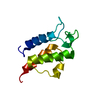
| ||||||||
|---|---|---|---|---|---|---|---|---|---|
| 1 |
| ||||||||
| Unit cell |
|
-Components
| #1: Protein | Mass: 12159.547 Da / Num. of mol.: 1 Source method: isolated from a genetically manipulated source Source: (gene. exp.)  |
|---|---|
| #2: Water | ChemComp-HOH /  Mass: 18.015 Da / Num. of mol.: 82 / Source method: isolated from a natural source / Formula: H2O Mass: 18.015 Da / Num. of mol.: 82 / Source method: isolated from a natural source / Formula: H2O |
| Sequence details | THE SEQUENCE OF THIS PROTEIN WAS NOT AVAILABLE AT THE UNIPROT KNOWLEDGEBASE DATABASE (UNIPROTKB) AT ...THE SEQUENCE OF THIS PROTEIN WAS NOT AVAILABLE AT THE UNIPROT KNOWLEDGEB |
-Experimental details
-Experiment
| Experiment | Method: X-RAY DIFFRACTION / Number of used crystals: 1 |
|---|
- Sample preparation
| Crystal | Density Matthews: 1.89 Å3/Da / Density % sol: 35.09 % |
|---|---|
| Crystal grow | Temperature: 293 K / Method: vapor diffusion, sitting drop / pH: 4.5 Details: 1.5 M NH4SO4, 0.2 M K,Na Tartrate, 0.1 M Na Acetate pH 4.5, VAPOR DIFFUSION, SITTING DROP, temperature 293K |
-Data collection
| Diffraction | Mean temperature: 100 K |
|---|---|
| Diffraction source | Source: ROTATING ANODE / Type: RIGAKU FR-E+ SUPERBRIGHT / Wavelength: 1.5418 Å |
| Detector | Type: RIGAKU RAXIS IV / Detector: IMAGE PLATE / Date: Jan 10, 2008 |
| Radiation | Protocol: SINGLE WAVELENGTH / Monochromatic (M) / Laue (L): M / Scattering type: x-ray |
| Radiation wavelength | Wavelength: 1.5418 Å / Relative weight: 1 |
| Reflection | Resolution: 1.64→50 Å / Num. all: 11276 / Num. obs: 11276 / % possible obs: 99.3 % / Observed criterion σ(F): 0 / Observed criterion σ(I): 0 / Redundancy: 9.7 % / Biso Wilson estimate: 23.5 Å2 / Rmerge(I) obs: 0.052 / Rsym value: 0.056 / Χ2: 1.973 / Net I/σ(I): 21.5 |
| Reflection shell | Resolution: 1.64→1.7 Å / Redundancy: 4.1 % / Rmerge(I) obs: 0.198 / Mean I/σ(I) obs: 6.18 / Num. unique all: 1054 / Rsym value: 0.183 / Χ2: 0.827 / % possible all: 93.2 |
-Phasing
| Phasing | Method: molecular replacement | |||||||||
|---|---|---|---|---|---|---|---|---|---|---|
| Phasing MR | Model details: Phaser MODE: MR_AUTO
|
- Processing
| Software |
| ||||||||||||||||||||||||||||||||||||||||||||||||||||||||||||||||||||||||||||||||||||||||||
|---|---|---|---|---|---|---|---|---|---|---|---|---|---|---|---|---|---|---|---|---|---|---|---|---|---|---|---|---|---|---|---|---|---|---|---|---|---|---|---|---|---|---|---|---|---|---|---|---|---|---|---|---|---|---|---|---|---|---|---|---|---|---|---|---|---|---|---|---|---|---|---|---|---|---|---|---|---|---|---|---|---|---|---|---|---|---|---|---|---|---|---|
| Refinement | Method to determine structure: MOLECULAR REPLACEMENT Starting model: PDB entry 1L0I Resolution: 1.64→32.34 Å / Cor.coef. Fo:Fc: 0.961 / Cor.coef. Fo:Fc free: 0.941 / SU B: 1.502 / SU ML: 0.055 / Cross valid method: THROUGHOUT / σ(F): 0 / σ(I): 0 / ESU R: 0.103 / ESU R Free: 0.105 / Stereochemistry target values: MAXIMUM LIKELIHOOD / Details: HYDROGENS HAVE BEEN ADDED IN THE RIDING POSITIONS
| ||||||||||||||||||||||||||||||||||||||||||||||||||||||||||||||||||||||||||||||||||||||||||
| Solvent computation | Ion probe radii: 0.8 Å / Shrinkage radii: 0.8 Å / VDW probe radii: 1.4 Å / Solvent model: MASK | ||||||||||||||||||||||||||||||||||||||||||||||||||||||||||||||||||||||||||||||||||||||||||
| Displacement parameters | Biso mean: 23.535 Å2
| ||||||||||||||||||||||||||||||||||||||||||||||||||||||||||||||||||||||||||||||||||||||||||
| Refinement step | Cycle: LAST / Resolution: 1.64→32.34 Å
| ||||||||||||||||||||||||||||||||||||||||||||||||||||||||||||||||||||||||||||||||||||||||||
| Refine LS restraints |
| ||||||||||||||||||||||||||||||||||||||||||||||||||||||||||||||||||||||||||||||||||||||||||
| LS refinement shell | Resolution: 1.64→1.68 Å / Total num. of bins used: 20
|
