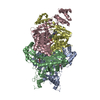
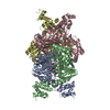
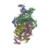
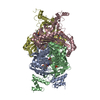
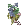
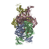
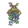
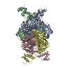
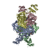
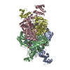
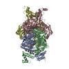
A: 3-hydroxy-3-methylglutaryl-coenzyme A reductase B: 3-hydroxy-3-methylglutaryl-coenzyme A reductase C: 3-hydroxy-3-methylglutaryl-coenzyme A reductase D: 3-hydroxy-3-methylglutaryl-coenzyme A reductase hetero molecules
Type: MAR CCD 165 mm / Detector: CCD / Date: Jul 10, 2003
Radiation
Protocol: SINGLE WAVELENGTH / Monochromatic (M) / Laue (L): M / Scattering type: x-ray
Radiation wavelength
Wavelength: 1 Å / Relative weight: 1
Reflection
Redundancy: 4.4 % / Av σ(I) over netI: 10.4 / Number: 279766 / Rmerge(I) obs: 0.06 / Χ2: 1 / D res high: 2.4 Å / D res low: 50 Å / Num. obs: 64140 / % possible obs: 97.9
Diffraction reflection shell
Highest resolution (Å)
Lowest resolution (Å)
% possible obs (%)
ID
Rmerge(I) obs
Chi squared
Redundancy
5.17
50
99.9
1
0.029
0.973
4.6
4.1
5.17
100
1
0.037
1.214
4.6
3.58
4.1
100
1
0.048
1.212
4.5
3.26
3.58
100
1
0.064
1.117
4.5
3.02
3.26
100
1
0.086
0.997
4.5
2.85
3.02
100
1
0.123
0.959
4.5
2.7
2.85
100
1
0.165
0.886
4.4
2.59
2.7
99
1
0.193
0.855
4.2
2.49
2.59
93.6
1
0.222
0.825
4
2.4
2.49
86.9
1
0.252
0.806
3.8
Reflection
Resolution: 2.4→50 Å / Num. obs: 64140 / % possible obs: 97.9 % / Redundancy: 4.4 % / Rmerge(I) obs: 0.06 / Χ2: 0.996 / Net I/σ(I): 10.4
Reflection shell
Resolution (Å)
Redundancy (%)
Rmerge(I) obs
Num. unique all
Χ2
% possible all
2.4-2.49
3.8
0.252
5669
0.806
86.9
2.49-2.59
4
0.222
6085
0.825
93.6
2.59-2.7
4.2
0.193
6488
0.855
99
2.7-2.85
4.4
0.165
6553
0.886
100
2.85-3.02
4.5
0.123
6493
0.959
100
3.02-3.26
4.5
0.086
6545
0.997
100
3.26-3.58
4.5
0.064
6551
1.117
100
3.58-4.1
4.5
0.048
6542
1.212
100
4.1-5.17
4.6
0.037
6583
1.214
100
5.17-50
4.6
0.029
6631
0.973
99.9
-
Phasing
Phasing
Method: molecular replacement
-
Processing
Software
Name
Version
Classification
NB
DENZO
datareduction
SCALEPACK
datascaling
MOLREP
phasing
REFMAC
refinement
PDB_EXTRACT
3.004
dataextraction
Refinement
Method to determine structure: MOLECULAR REPLACEMENT / Resolution: 2.4→50 Å / Cor.coef. Fo:Fc: 0.947 / Cor.coef. Fo:Fc free: 0.915 / SU B: 8.087 / SU ML: 0.187 / Cross valid method: THROUGHOUT / σ(F): 0 / ESU R: 0.581 / ESU R Free: 0.272 / Stereochemistry target values: MAXIMUM LIKELIHOOD / Details: HYDROGENS HAVE BEEN ADDED IN THE RIDING POSITIONS
Rfactor
Num. reflection
% reflection
Selection details
Rfree
0.238
3290
5.1 %
RANDOM
Rwork
0.19
-
-
-
obs
0.192
64058
97.78 %
-
Solvent computation
Ion probe radii: 0.8 Å / Shrinkage radii: 0.8 Å / VDW probe radii: 1.4 Å / Solvent model: BABINET MODEL WITH MASK
Displacement parameters
Biso mean: 34.367 Å2
Baniso -1
Baniso -2
Baniso -3
1-
3.4 Å2
0 Å2
1.36 Å2
2-
-
-1.99 Å2
0 Å2
3-
-
-
-0.1 Å2
Refinement step
Cycle: LAST / Resolution: 2.4→50 Å
Protein
Nucleic acid
Ligand
Solvent
Total
Num. atoms
12282
0
148
645
13075
Refine LS restraints
Refine-ID
Type
Dev ideal
Dev ideal target
Number
X-RAY DIFFRACTION
r_bond_refined_d
0.008
0.021
12618
X-RAY DIFFRACTION
r_bond_other_d
0.002
0.02
11691
X-RAY DIFFRACTION
r_angle_refined_deg
1.083
1.983
17057
X-RAY DIFFRACTION
r_angle_other_deg
0.755
3
27238
X-RAY DIFFRACTION
r_dihedral_angle_1_deg
5.564
5
1646
X-RAY DIFFRACTION
r_chiral_restr
0.061
0.2
1955
X-RAY DIFFRACTION
r_gen_planes_refined
0.003
0.02
14114
X-RAY DIFFRACTION
r_gen_planes_other
0.002
0.02
2345
X-RAY DIFFRACTION
r_nbd_refined
0.177
0.2
2723
X-RAY DIFFRACTION
r_nbd_other
0.211
0.2
14024
X-RAY DIFFRACTION
r_nbtor_other
0.08
0.2
7679
X-RAY DIFFRACTION
r_xyhbond_nbd_refined
0.15
0.2
649
X-RAY DIFFRACTION
r_symmetry_vdw_refined
0.114
0.2
14
X-RAY DIFFRACTION
r_symmetry_vdw_other
0.187
0.2
46
X-RAY DIFFRACTION
r_symmetry_hbond_refined
0.144
0.2
4
X-RAY DIFFRACTION
r_mcbond_it
0.383
1.5
8166
X-RAY DIFFRACTION
r_mcangle_it
0.734
2
13054
X-RAY DIFFRACTION
r_scbond_it
1.03
3
4452
X-RAY DIFFRACTION
r_scangle_it
1.852
4.5
4003
LS refinement shell
Resolution: 2.4→2.462 Å / Total num. of bins used: 20
Rfactor
Num. reflection
Rfree
0.287
210
Rwork
0.232
3873
all
-
4083
+
About Yorodumi
-
News
-
Feb 9, 2022. New format data for meta-information of EMDB entries
New format data for meta-information of EMDB entries
Version 3 of the EMDB header file is now the official format.
The previous official version 1.9 will be removed from the archive.
In the structure databanks used in Yorodumi, some data are registered as the other names, "COVID-19 virus" and "2019-nCoV". Here are the details of the virus and the list of structure data.
Jan 31, 2019. EMDB accession codes are about to change! (news from PDBe EMDB page)
EMDB accession codes are about to change! (news from PDBe EMDB page)
The allocation of 4 digits for EMDB accession codes will soon come to an end. Whilst these codes will remain in use, new EMDB accession codes will include an additional digit and will expand incrementally as the available range of codes is exhausted. The current 4-digit format prefixed with “EMD-” (i.e. EMD-XXXX) will advance to a 5-digit format (i.e. EMD-XXXXX), and so on. It is currently estimated that the 4-digit codes will be depleted around Spring 2019, at which point the 5-digit format will come into force.
The EM Navigator/Yorodumi systems omit the EMD- prefix.
Related info.:Q: What is EMD? / ID/Accession-code notation in Yorodumi/EM Navigator
Yorodumi is a browser for structure data from EMDB, PDB, SASBDB, etc.
This page is also the successor to EM Navigator detail page, and also detail information page/front-end page for Omokage search.
The word "yorodu" (or yorozu) is an old Japanese word meaning "ten thousand". "mi" (miru) is to see.
Related info.:EMDB / PDB / SASBDB / Comparison of 3 databanks / Yorodumi Search / Aug 31, 2016. New EM Navigator & Yorodumi / Yorodumi Papers / Jmol/JSmol / Function and homology information / Changes in new EM Navigator and Yorodumi
 Movie
Movie Controller

 Yorodumi
 Open data
 Basic information
 Components
 Keywords
 Function and homology information
 Homo sapiens (human)
 X-RAY DIFFRACTION /
  Authors
 Citation
 Structure visualization
 Downloads & links
 Other downloads


 PDBj
PDBj

 Assembly
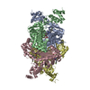

 Mass: 515.549 Da / Num. of mol.: 4 / Source method: obtained synthetically / Formula: C27H31F2N3O5
 Mass: 18.015 Da / Num. of mol.: 645 / Source method: isolated from a natural source / Formula: H2O
 Sample preparation
 / Beamline: 17-BM / Wavelength: 1 Å
 Processing