 Movie
Movie Controller

[English] 日本語
 Yorodumi
- PDB-3ccz: Thermodynamic and structure guided design of statin hmg-coa reduc... -
+ Open data

- Basic information
| Entry | Database: PDB / ID: 3ccz | ||||||
|---|---|---|---|---|---|---|---|
| Title | Thermodynamic and structure guided design of statin hmg-coa reductase inhibitors | ||||||
 Components Components | 3-hydroxy-3-methylglutaryl-coenzyme A reductase | ||||||
 Keywords Keywords | OXIDOREDUCTASE / CHOLESTEROL BIOSYNTHESIS / HMG-COA / NADPH / STATIN / Alternative splicing / Endoplasmic reticulum / Glycoprotein / Lipid synthesis / Membrane / Peroxisome / Polymorphism / Steroid biosynthesis / Transmembrane | ||||||
| Function / homology |  Function and homology information Function and homology informationhydroxymethylglutaryl-CoA reductase (NADPH) / hydroxymethylglutaryl-CoA reductase (NADPH) activity / sterol biosynthetic process / isopentenyl diphosphate biosynthetic process, mevalonate pathway / GTPase regulator activity / negative regulation of amyloid-beta clearance / coenzyme A binding / farnesyl diphosphate biosynthetic process, mevalonate pathway / zymosterol biosynthetic process / : ...hydroxymethylglutaryl-CoA reductase (NADPH) / hydroxymethylglutaryl-CoA reductase (NADPH) activity / sterol biosynthetic process / isopentenyl diphosphate biosynthetic process, mevalonate pathway / GTPase regulator activity / negative regulation of amyloid-beta clearance / coenzyme A binding / farnesyl diphosphate biosynthetic process, mevalonate pathway / zymosterol biosynthetic process / : / : / Lanosterol biosynthesis / geranylgeranyl diphosphate biosynthetic process / coenzyme A metabolic process / EGR2 and SOX10-mediated initiation of Schwann cell myelination / isoprenoid biosynthetic process / cholesterol biosynthetic process / peroxisomal membrane / cytoplasmic side of endoplasmic reticulum membrane / negative regulation of protein secretion / NADPH binding / Activation of gene expression by SREBF (SREBP) / negative regulation of protein catabolic process / PPARA activates gene expression / endoplasmic reticulum membrane / endoplasmic reticulum Similarity search - Function | ||||||
| Biological species |  Homo sapiens (human) Homo sapiens (human) | ||||||
| Method |  X-RAY DIFFRACTION / SYNCHROTRON / MOLECULAR REPLACEMENT / molecular replacement / Resolution: 1.7 Å X-RAY DIFFRACTION / SYNCHROTRON / MOLECULAR REPLACEMENT / molecular replacement / Resolution: 1.7 Å | ||||||
 Authors Authors | Pavlovsky, A. / Sarver, R.W. / Harris, M.S. / Finzel, B.C. | ||||||
 Citation Citation | Journal: J.Med.Chem. / Year: 2008 Title: Thermodynamic and structure guided design of statin based inhibitors of 3-hydroxy-3-methylglutaryl coenzyme a reductase. Authors: Sarver, R.W. / Bills, E. / Bolton, G. / Bratton, L.D. / Caspers, N.L. / Dunbar, J.B. / Harris, M.S. / Hutchings, R.H. / Kennedy, R.M. / Larsen, S.D. / Pavlovsky, A. / Pfefferkorn, J.A. / Bainbridge, G. | ||||||
| History |
|
- Structure visualization
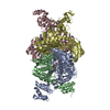
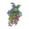
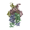
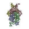
| Structure viewer | Molecule: MolmilJmol/JSmol |
|---|
- Downloads & links
-Download
| PDBx/mmCIF format | 3ccz.cif.gz | 331.9 KB | Display | PDBx/mmCIF format |
|---|---|---|---|---|
| PDB format | pdb3ccz.ent.gz | 269 KB | Display | PDB format |
| PDBx/mmJSON format | 3ccz.json.gz | Tree view | PDBx/mmJSON format | |
| Others |  Other downloads Other downloads |
-Validation report
| Arichive directory | https://data.pdbj.org/pub/pdb/validation_reports/cc/3cczftp://data.pdbj.org/pub/pdb/validation_reports/cc/3ccz | HTTPS FTP |
|---|
-Related structure data
| Related structure data |  3cctC  3ccwC  3cd0C  3cd5C 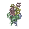 3cd7C  3cdaC 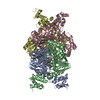 3cdbC C: citing same article ( |
|---|---|
| Similar structure data |


-Links
 PDBj
PDBj

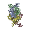
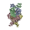
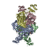
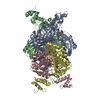
- Assembly
| Deposited unit | 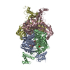
| ||||||||
|---|---|---|---|---|---|---|---|---|---|
| 1 |
| ||||||||
| Unit cell |
|
-Components
| #1: Protein | Mass: 47479.551 Da / Num. of mol.: 4 / Fragment: CATALYTIC DOMAIN (RESIDUES 441-875) / Mutation: M485I Source method: isolated from a genetically manipulated source Source: (gene. exp.) Homo sapiens (human) / Gene: HMGCR / Production host:  References: UniProt: P04035, hydroxymethylglutaryl-CoA reductase (NADPH) #2: Chemical | ChemComp-SO4 /   Mass: 96.063 Da / Num. of mol.: 4 / Source method: obtained synthetically / Formula: SO4 Mass: 96.063 Da / Num. of mol.: 4 / Source method: obtained synthetically / Formula: SO4#3: Chemical | ChemComp-5HI / ( 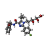  Mass: 527.584 Da / Num. of mol.: 4 / Source method: obtained synthetically / Formula: C28H34FN3O6 Mass: 527.584 Da / Num. of mol.: 4 / Source method: obtained synthetically / Formula: C28H34FN3O6#4: Water | ChemComp-HOH / |  Mass: 18.015 Da / Num. of mol.: 1236 / Source method: isolated from a natural source / Formula: H2O Mass: 18.015 Da / Num. of mol.: 1236 / Source method: isolated from a natural source / Formula: H2O |
|---|
-Experimental details
-Experiment
| Experiment | Method: X-RAY DIFFRACTION / Number of used crystals: 1 |
|---|
- Sample preparation
| Crystal | Density Matthews: 2.44 Å3/Da / Density % sol: 49.62 % |
|---|---|
| Crystal grow | Temperature: 293 K / Method: vapor diffusion, hanging drop / pH: 8 Details: protein 15-20 mg/ml, Ligand (saturated),PEG 4000, MgCl2 0.2M, Tris-HCL pH8 0.1M, 7-10 days, pH 8.0, VAPOR DIFFUSION, HANGING DROP, temperature 293K |
-Data collection
| Diffraction | Mean temperature: 100 K | |||||||||||||||||||||||||||||||||||||||||||||||||||||||||||||||||||||||||||||
|---|---|---|---|---|---|---|---|---|---|---|---|---|---|---|---|---|---|---|---|---|---|---|---|---|---|---|---|---|---|---|---|---|---|---|---|---|---|---|---|---|---|---|---|---|---|---|---|---|---|---|---|---|---|---|---|---|---|---|---|---|---|---|---|---|---|---|---|---|---|---|---|---|---|---|---|---|---|---|
| Diffraction source | Source: SYNCHROTRON / Site: APS  / Beamline: 17-ID / Wavelength: 1 Å / Beamline: 17-ID / Wavelength: 1 Å | |||||||||||||||||||||||||||||||||||||||||||||||||||||||||||||||||||||||||||||
| Detector | Type: ADSC QUANTUM 210 / Detector: CCD / Date: Jun 9, 2004 | |||||||||||||||||||||||||||||||||||||||||||||||||||||||||||||||||||||||||||||
| Radiation | Protocol: SINGLE WAVELENGTH / Monochromatic (M) / Laue (L): M / Scattering type: x-ray | |||||||||||||||||||||||||||||||||||||||||||||||||||||||||||||||||||||||||||||
| Radiation wavelength | Wavelength: 1 Å / Relative weight: 1 | |||||||||||||||||||||||||||||||||||||||||||||||||||||||||||||||||||||||||||||
| Reflection | Redundancy: 3.7 % / Av σ(I) over netI: 26.4 / Number: 694736 / Rmerge(I) obs: 0.041 / Χ2: 0.85 / D res high: 1.7 Å / D res low: 50 Å / Num. obs: 187138 / % possible obs: 93.6 | |||||||||||||||||||||||||||||||||||||||||||||||||||||||||||||||||||||||||||||
| Diffraction reflection shell |
| |||||||||||||||||||||||||||||||||||||||||||||||||||||||||||||||||||||||||||||
| Reflection | Resolution: 1.7→50 Å / Num. obs: 187138 / % possible obs: 93.6 % / Redundancy: 3.7 % / Rmerge(I) obs: 0.041 / Χ2: 0.845 / Net I/σ(I): 26.4 | |||||||||||||||||||||||||||||||||||||||||||||||||||||||||||||||||||||||||||||
| Reflection shell |
|
-Phasing
| Phasing | Method: molecular replacement |
|---|
- Processing
| Software |
| ||||||||||||||||||||||||||||||||||||||||||||||||||||||||||||||||||||||||||||||||||||||||||||||||||||
|---|---|---|---|---|---|---|---|---|---|---|---|---|---|---|---|---|---|---|---|---|---|---|---|---|---|---|---|---|---|---|---|---|---|---|---|---|---|---|---|---|---|---|---|---|---|---|---|---|---|---|---|---|---|---|---|---|---|---|---|---|---|---|---|---|---|---|---|---|---|---|---|---|---|---|---|---|---|---|---|---|---|---|---|---|---|---|---|---|---|---|---|---|---|---|---|---|---|---|---|---|---|
| Refinement | Method to determine structure: MOLECULAR REPLACEMENT / Resolution: 1.7→50 Å / Cor.coef. Fo:Fc: 0.935 / Cor.coef. Fo:Fc free: 0.92 / SU B: 2.616 / SU ML: 0.086 / Cross valid method: THROUGHOUT / σ(F): 0 / ESU R: 0.131 / ESU R Free: 0.122 / Stereochemistry target values: MAXIMUM LIKELIHOOD / Details: HYDROGENS HAVE BEEN ADDED IN THE RIDING POSITIONS
| ||||||||||||||||||||||||||||||||||||||||||||||||||||||||||||||||||||||||||||||||||||||||||||||||||||
| Solvent computation | Ion probe radii: 0.8 Å / Shrinkage radii: 0.8 Å / VDW probe radii: 1.4 Å / Solvent model: BABINET MODEL WITH MASK | ||||||||||||||||||||||||||||||||||||||||||||||||||||||||||||||||||||||||||||||||||||||||||||||||||||
| Displacement parameters | Biso mean: 25.234 Å2
| ||||||||||||||||||||||||||||||||||||||||||||||||||||||||||||||||||||||||||||||||||||||||||||||||||||
| Refinement step | Cycle: LAST / Resolution: 1.7→50 Å
| ||||||||||||||||||||||||||||||||||||||||||||||||||||||||||||||||||||||||||||||||||||||||||||||||||||
| Refine LS restraints |
| ||||||||||||||||||||||||||||||||||||||||||||||||||||||||||||||||||||||||||||||||||||||||||||||||||||
| LS refinement shell | Resolution: 1.699→1.743 Å / Total num. of bins used: 20
|
