 Movie
Movie Controller


+ Open data

- Basic information
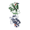
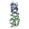
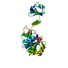
| Entry | Database: PDB / ID: 3c9g | ||||||
|---|---|---|---|---|---|---|---|
| Title | Crystal structure of uncharacterized UPF0201 protein AF_135 | ||||||
 Components Components | UPF0200/UPF0201 protein AF_1395 | ||||||
 Keywords Keywords | NUCLEOTIDE BINDING PROTEIN / uncharacterized UPF0201 protein AF_135 / Structural genomics / PSI-II / dimer / Protein Structure Initiative / New York SGX Research Center for Structural Genomics / NYSGXRC / ATP-binding / Nucleotide-binding | ||||||
| Function / homology |  Function and homology information Function and homology information | ||||||
| Biological species |   Archaeoglobus fulgidus DSM 4304 (archaea) Archaeoglobus fulgidus DSM 4304 (archaea) | ||||||
| Method |  X-RAY DIFFRACTION / SYNCHROTRON / MOLECULAR REPLACEMENT / Resolution: 2.3 Å X-RAY DIFFRACTION / SYNCHROTRON / MOLECULAR REPLACEMENT / Resolution: 2.3 Å | ||||||
 Authors Authors | Sugadev, R. / Ozyurt, S. / Burley, S.K. / Swaminathan, S. / New York SGX Research Center for Structural Genomics (NYSGXRC) | ||||||
 Citation Citation | Journal: To be Published Title: Crystal structure of uncharacterized UPF0201 protein AF_135. Authors: Sugadev, R. / Ozyurt, S. / Burley, S.K. / Swaminathan, S. | ||||||
| History |
|
- Structure visualization
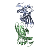
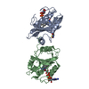
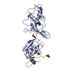
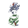
| Structure viewer | Molecule: MolmilJmol/JSmol |
|---|
- Downloads & links
-Download
| PDBx/mmCIF format | 3c9g.cif.gz | 63.5 KB | Display | PDBx/mmCIF format |
|---|---|---|---|---|
| PDB format | pdb3c9g.ent.gz | 47.1 KB | Display | PDB format |
| PDBx/mmJSON format | 3c9g.json.gz | Tree view | PDBx/mmJSON format | |
| Others |  Other downloads Other downloads |
-Validation report
| Arichive directory | https://data.pdbj.org/pub/pdb/validation_reports/c9/3c9gftp://data.pdbj.org/pub/pdb/validation_reports/c9/3c9g | HTTPS FTP |
|---|
-Related structure data
| Related structure data |  2nwuS S: Starting model for refinement |
|---|---|
| Similar structure data | |
| Other databases |


-Links
 PDBj
PDBj- Assembly
| Deposited unit | 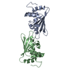
| ||||||||
|---|---|---|---|---|---|---|---|---|---|
| 1 |
| ||||||||
| Unit cell |
|
-Components
| #1: Protein | Mass: 16447.885 Da / Num. of mol.: 2 / Fragment: Residues 183-323 Source method: isolated from a genetically manipulated source Source: (gene. exp.) Archaeoglobus fulgidus DSM 4304 (archaea)Species: Archaeoglobus fulgidus / Strain: DSM 4304 / VC-16 / JCM 9628 / NBRC 100126 / Gene: AF_1395 / Plasmid: pSGX3(BC) / Species (production host): Escherichia coli / Production host:  #2: Water | ChemComp-HOH / |  Mass: 18.015 Da / Num. of mol.: 20 / Source method: isolated from a natural source / Formula: H2O Mass: 18.015 Da / Num. of mol.: 20 / Source method: isolated from a natural source / Formula: H2O |
|---|
-Experimental details
-Experiment
| Experiment | Method: X-RAY DIFFRACTION / Number of used crystals: 1 |
|---|
- Sample preparation
| Crystal | Density Matthews: 2.32 Å3/Da / Density % sol: 46.95 % Description: The structure factor file contains Friedel pairs |
|---|---|
| Crystal grow | Temperature: 298 K / Method: vapor diffusion, sitting drop / pH: 5.2 Details: 0.1 M Sodium citrate pH 5.2, 30% PEG 4000, 0.1M Ammonium sulfate, VAPOR DIFFUSION, SITTING DROP, temperature 298K |
-Data collection
| Diffraction | Mean temperature: 100 K |
|---|---|
| Diffraction source | Source: SYNCHROTRON / Site: APS  / Beamline: 31-ID / Wavelength: 0.9795 Å / Beamline: 31-ID / Wavelength: 0.9795 Å |
| Detector | Type: MAR CCD 165 mm / Detector: CCD / Date: Jun 3, 2006 / Details: SGX_CAT |
| Radiation | Monochromator: SGX_CAT / Protocol: SINGLE WAVELENGTH / Monochromatic (M) / Laue (L): M / Scattering type: x-ray |
| Radiation wavelength | Wavelength: 0.9795 Å / Relative weight: 1 |
| Reflection | Resolution: 2.29→50 Å / Num. all: 25121 / Num. obs: 25121 / % possible obs: 99.3 % / Observed criterion σ(F): 0 / Observed criterion σ(I): 0 / Redundancy: 13.7 % / Biso Wilson estimate: 30.4 Å2 / Rmerge(I) obs: 0.098 / Net I/σ(I): 6.5 |
- Processing
| Software |
| ||||||||||||||||||||
|---|---|---|---|---|---|---|---|---|---|---|---|---|---|---|---|---|---|---|---|---|---|
| Refinement | Method to determine structure: MOLECULAR REPLACEMENT Starting model: PDB entry 2NWU Resolution: 2.3→24.45 Å / Rfactor Rfree error: 0.01 / Data cutoff high absF: 147025.62 / Data cutoff low absF: 0 / Isotropic thermal model: RESTRAINED / Cross valid method: THROUGHOUT / σ(F): 2 / Stereochemistry target values: Engh & Huber Details: The Friedel pairs were used in phasing. Residues listed as missing in Remark 465 are due to lack of electron density. Residues with missing atoms listed in Remark 470 are due to lack of ...Details: The Friedel pairs were used in phasing. Residues listed as missing in Remark 465 are due to lack of electron density. Residues with missing atoms listed in Remark 470 are due to lack of electron density for side chains and modeled as alanines. The R free value is slightly higher.
| ||||||||||||||||||||
| Solvent computation | Solvent model: FLAT MODEL / Bsol: 31.7094 Å2 / ksol: 0.315086 e/Å3 | ||||||||||||||||||||
| Displacement parameters | Biso mean: 45.8 Å2
| ||||||||||||||||||||
| Refine analyze |
| ||||||||||||||||||||
| Refinement step | Cycle: LAST / Resolution: 2.3→24.45 Å
| ||||||||||||||||||||
| Refine LS restraints |
| ||||||||||||||||||||
| LS refinement shell | Resolution: 2.3→2.44 Å / Rfactor Rfree error: 0.034 / Total num. of bins used: 6
| ||||||||||||||||||||
| Xplor file |
|
