 Movie
Movie Controller


+ Open data

- Basic information
| Entry | Database: PDB / ID: 3c8p | ||||||
|---|---|---|---|---|---|---|---|
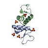
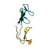
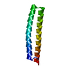
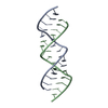
| Title | X-ray structure of Viscotoxin A1 from Viscum album L. | ||||||
 Components Components | Viscotoxin A1 | ||||||
 Keywords Keywords | TOXIN / helix turn helix | ||||||
| Function / homology |  Function and homology information Function and homology information | ||||||
| Biological species |  Viscum album (European mistletoe) Viscum album (European mistletoe) | ||||||
| Method |  X-RAY DIFFRACTION / SYNCHROTRON / SAD / Resolution: 1.25 Å X-RAY DIFFRACTION / SYNCHROTRON / SAD / Resolution: 1.25 Å | ||||||
 Authors Authors | Pal, A. / Debreczeni, J.E. / Sevvana, M. / Gruene, T. / Kahle, B. / Zeeck, A. / Sheldrick, G.M. | ||||||
 Citation Citation | Journal: Acta Crystallogr.,Sect.D / Year: 2008 Title: Structures of viscotoxins A1 and B2 from European mistletoe solved using native data alone Authors: Pal, A. / Debreczeni, J.E. / Sevvana, M. / Gruene, T. / Kahle, B. / Zeeck, A. / Sheldrick, G.M. | ||||||
| History |
|
- Structure visualization
| Structure viewer | Molecule: MolmilJmol/JSmol |
|---|
- Downloads & links
-Download
| PDBx/mmCIF format | 3c8p.cif.gz | 49.6 KB | Display | PDBx/mmCIF format |
|---|---|---|---|---|
| PDB format | pdb3c8p.ent.gz | 36.6 KB | Display | PDB format |
| PDBx/mmJSON format | 3c8p.json.gz | Tree view | PDBx/mmJSON format | |
| Others |  Other downloads Other downloads |
-Validation report
| Arichive directory | https://data.pdbj.org/pub/pdb/validation_reports/c8/3c8pftp://data.pdbj.org/pub/pdb/validation_reports/c8/3c8p | HTTPS FTP |
|---|
-Related structure data


-Links
 PDBj
PDBj
- Assembly
| Deposited unit | 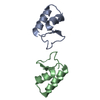
| |||||||||
|---|---|---|---|---|---|---|---|---|---|---|
| 1 |
| |||||||||
| Unit cell |
| |||||||||
| Components on special symmetry positions |
|
-Components
| #1: Protein/peptide | Mass: 4896.630 Da / Num. of mol.: 2 / Source method: isolated from a natural source / Source: (natural) Viscum album (European mistletoe) / References: UniProt: D0VWT3*PLUS#2: Water | ChemComp-HOH / |  Mass: 18.015 Da / Num. of mol.: 135 / Source method: isolated from a natural source / Formula: H2O Mass: 18.015 Da / Num. of mol.: 135 / Source method: isolated from a natural source / Formula: H2OHas protein modification | Y | |
|---|
-Experimental details
-Experiment
| Experiment | Method: X-RAY DIFFRACTION / Number of used crystals: 1 |
|---|
- Sample preparation
| Crystal | Density Matthews: 2.6 Å3/Da / Density % sol: 52.7 % |
|---|---|
| Crystal grow | Temperature: 298 K / pH: 7.5 Details: 1.4M Sodium citrate, 0.1 M HEPES pH 7.5, vapor diffusion, temperature 298K, pH 7.50 |
-Data collection
| Diffraction |
| ||||||||||||||||||
|---|---|---|---|---|---|---|---|---|---|---|---|---|---|---|---|---|---|---|---|
| Diffraction source |
| ||||||||||||||||||
| Detector |
| ||||||||||||||||||
| Radiation |
| ||||||||||||||||||
| Radiation wavelength |
| ||||||||||||||||||
| Reflection | Resolution: 1.25→47.16 Å / Num. obs: 29217 / % possible obs: 100 % / Observed criterion σ(I): 0 / Redundancy: 1.8 % / Biso Wilson estimate: 11.6 Å2 / Rmerge(I) obs: 0.0891 / Rsym value: 0.031 / Net I/σ(I): 25.5 | ||||||||||||||||||
| Reflection shell | Resolution: 1.25→1.35 Å / Redundancy: 1.75 % / Rmerge(I) obs: 0.3038 / Mean I/σ(I) obs: 2.61 / Rsym value: 0.3038 / % possible all: 100 |

- Processing
| Software |
| ||||||||||||||||||||||||||||||||||||
|---|---|---|---|---|---|---|---|---|---|---|---|---|---|---|---|---|---|---|---|---|---|---|---|---|---|---|---|---|---|---|---|---|---|---|---|---|---|
| Refinement | Method to determine structure: SAD / Resolution: 1.25→47.16 Å / Num. parameters: 7269 / Num. restraintsaints: 8599 / Cross valid method: FREE R / σ(F): 0 / Stereochemistry target values: ENGH AND HUBER
| ||||||||||||||||||||||||||||||||||||
| Solvent computation | Solvent model: MEWS AND KRETSINGER | ||||||||||||||||||||||||||||||||||||
| Displacement parameters | Biso mean: 17.81 Å2 | ||||||||||||||||||||||||||||||||||||
| Refine analyze | Num. disordered residues: 1 / Occupancy sum non hydrogen: 801.79 | ||||||||||||||||||||||||||||||||||||
| Refinement step | Cycle: LAST / Resolution: 1.25→47.16 Å
| ||||||||||||||||||||||||||||||||||||
| Refine LS restraints |
|
