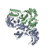
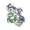
 Movie
Movie Controller

[English] 日本語
 Yorodumi
- PDB-3c6u: Crystal Structure of HIV Reverse Transcriptase in complex with in... -
+ Open data

- Basic information
| Entry | Database: PDB / ID: 3c6u | ||||||
|---|---|---|---|---|---|---|---|
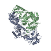
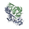
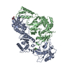
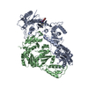
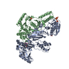
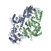
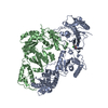
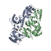
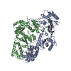
| Title | Crystal Structure of HIV Reverse Transcriptase in complex with inhibitor 22 | ||||||
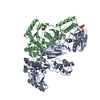
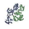
 Components Components | (Reverse transcriptase) x 2 | ||||||
 Keywords Keywords | TRANSFERASE / HIV-1 reverse transcriptase / non-nucleoside inhibition / nucleotidyltrasferase / AIDS / Aspartyl protease / Capsid maturation / Capsid protein / Cytoplasm / DNA integration / DNA recombination / DNA-directed DNA polymerase / Endonuclease / Host-virus interaction / Hydrolase / Lipoprotein / Magnesium / Membrane / Metal-binding / Multifunctional enzyme / Myristate / Nuclease / Nucleotidyltransferase / Nucleus / Phosphoprotein / Protease / RNA-binding / RNA-directed DNA polymerase / Viral nucleoprotein / Virion / Zinc / Zinc-finger | ||||||
| Function / homology |  Function and homology information Function and homology informationintegrase activity / Integration of viral DNA into host genomic DNA / Autointegration results in viral DNA circles / Minus-strand DNA synthesis / Plus-strand DNA synthesis / Uncoating of the HIV Virion / 2-LTR circle formation / Vpr-mediated nuclear import of PICs / Early Phase of HIV Life Cycle / Integration of provirus ...integrase activity / Integration of viral DNA into host genomic DNA / Autointegration results in viral DNA circles / Minus-strand DNA synthesis / Plus-strand DNA synthesis / Uncoating of the HIV Virion / 2-LTR circle formation / Vpr-mediated nuclear import of PICs / Early Phase of HIV Life Cycle / Integration of provirus / APOBEC3G mediated resistance to HIV-1 infection / Binding and entry of HIV virion / viral life cycle / HIV-1 retropepsin / symbiont-mediated activation of host apoptosis / retroviral ribonuclease H / exoribonuclease H / exoribonuclease H activity / Assembly Of The HIV Virion / protein processing / viral genome integration into host DNA / Budding and maturation of HIV virion / establishment of integrated proviral latency / RNA-directed DNA polymerase / RNA stem-loop binding / viral penetration into host nucleus / host multivesicular body / RNA-directed DNA polymerase activity / RNA-DNA hybrid ribonuclease activity / Transferases; Transferring phosphorus-containing groups; Nucleotidyltransferases / peptidase activity / host cell / viral nucleocapsid / DNA recombination / DNA-directed DNA polymerase / aspartic-type endopeptidase activity / Hydrolases; Acting on ester bonds / DNA-directed DNA polymerase activity / symbiont-mediated suppression of host gene expression / viral translational frameshifting / symbiont entry into host cell / lipid binding / host cell nucleus / host cell plasma membrane / virion membrane / structural molecule activity / DNA binding / zinc ion binding / identical protein binding Similarity search - Function | ||||||
| Biological species |  HIV-1 M:B_HXB2R (virus) HIV-1 M:B_HXB2R (virus) | ||||||
| Method |  X-RAY DIFFRACTION / SYNCHROTRON / FOURIER SYNTHESIS / Resolution: 2.7 Å X-RAY DIFFRACTION / SYNCHROTRON / FOURIER SYNTHESIS / Resolution: 2.7 Å | ||||||
 Authors Authors | Yan, Y. / Prasad, S. | ||||||
 Citation Citation | Journal: Bioorg.Med.Chem.Lett. / Year: 2008 Title: The design and synthesis of diaryl ether second generation HIV-1 non-nucleoside reverse transcriptase inhibitors (NNRTIs) with enhanced potency versus key clinical mutations. Authors: Tucker, T.J. / Saggar, S. / Sisko, J.T. / Tynebor, R.M. / Williams, T.M. / Felock, P.J. / Flynn, J.A. / Lai, M.T. / Liang, Y. / McGaughey, G. / Liu, M. / Miller, M. / Moyer, G. / Munshi, V. ...Authors: Tucker, T.J. / Saggar, S. / Sisko, J.T. / Tynebor, R.M. / Williams, T.M. / Felock, P.J. / Flynn, J.A. / Lai, M.T. / Liang, Y. / McGaughey, G. / Liu, M. / Miller, M. / Moyer, G. / Munshi, V. / Perlow-Poehnelt, R. / Prasad, S. / Sanchez, R. / Torrent, M. / Vacca, J.P. / Wan, B.L. / Yan, Y. | ||||||
| History |
|
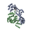
- Structure visualization
| Structure viewer | Molecule: MolmilJmol/JSmol |
|---|
- Downloads & links
-Download
| PDBx/mmCIF format | 3c6u.cif.gz | 216.4 KB | Display | PDBx/mmCIF format |
|---|---|---|---|---|
| PDB format | pdb3c6u.ent.gz | 171.2 KB | Display | PDB format |
| PDBx/mmJSON format | 3c6u.json.gz | Tree view | PDBx/mmJSON format | |
| Others |  Other downloads Other downloads |
-Validation report
| Arichive directory | https://data.pdbj.org/pub/pdb/validation_reports/c6/3c6uftp://data.pdbj.org/pub/pdb/validation_reports/c6/3c6u | HTTPS FTP |
|---|
-Related structure data
| Related structure data | 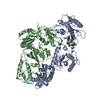 3c6tC 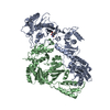 2rf2S C: citing same article ( S: Starting model for refinement |
|---|---|
| Similar structure data |


-Links
 PDBj
PDBj


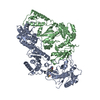
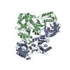
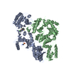
- Assembly
| Deposited unit | 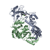
| ||||||||
|---|---|---|---|---|---|---|---|---|---|
| 1 |
| ||||||||
| Unit cell |
|
-Components
| #1: Protein | Mass: 64895.316 Da / Num. of mol.: 1 Source method: isolated from a genetically manipulated source Source: (gene. exp.) HIV-1 M:B_HXB2R (virus) / Genus: Lentivirus / Species: Human immunodeficiency virus 1 / Strain: HXB2 isolate / Gene: gag-pol / Production host:  References: UniProt: P04585, RNA-directed DNA polymerase, DNA-directed DNA polymerase |
|---|---|
| #2: Protein | Mass: 51731.418 Da / Num. of mol.: 1 Source method: isolated from a genetically manipulated source Source: (gene. exp.) HIV-1 M:B_HXB2R (virus) / Genus: Lentivirus / Species: Human immunodeficiency virus 1 / Strain: HXB2 isolate / Gene: gag-pol / Production host: |
| #3: Chemical | ChemComp-M22 / 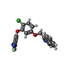  Mass: 410.253 Da / Num. of mol.: 1 / Source method: obtained synthetically / Formula: C21H13Cl2N3O2 Mass: 410.253 Da / Num. of mol.: 1 / Source method: obtained synthetically / Formula: C21H13Cl2N3O2 |
| #4: Water | ChemComp-HOH /  Mass: 18.015 Da / Num. of mol.: 375 / Source method: isolated from a natural source / Formula: H2O Mass: 18.015 Da / Num. of mol.: 375 / Source method: isolated from a natural source / Formula: H2O |
-Experimental details
-Experiment
| Experiment | Method: X-RAY DIFFRACTION / Number of used crystals: 1 |
|---|
- Sample preparation
| Crystal | Density Matthews: 3.06 Å3/Da / Density % sol: 59.86 % |
|---|---|
| Crystal grow | Temperature: 298 K / Method: vapor diffusion, sitting drop / pH: 6.1 Details: sodium citrate, pH 6.1, VAPOR DIFFUSION, SITTING DROP, temperature 298K |
-Data collection
| Diffraction | Mean temperature: 100 K |
|---|---|
| Diffraction source | Source: SYNCHROTRON / Site: APS  / Beamline: 17-ID / Wavelength: 1 Å / Beamline: 17-ID / Wavelength: 1 Å |
| Detector | Type: ADSC QUANTUM 210 / Detector: CCD / Date: Aug 9, 2005 |
| Radiation | Protocol: SINGLE WAVELENGTH / Monochromatic (M) / Laue (L): M / Scattering type: x-ray |
| Radiation wavelength | Wavelength: 1 Å / Relative weight: 1 |
| Reflection | Resolution: 2.7→50 Å / Num. all: 39464 / Num. obs: 35005 / % possible obs: 88.7 % / Observed criterion σ(F): 0 / Observed criterion σ(I): 0 / Redundancy: 4.6 % / Biso Wilson estimate: 73.676 Å2 / Rmerge(I) obs: 0.086 / Rsym value: 0.086 / Net I/σ(I): 12.6 |
| Reflection shell | Resolution: 2.7→2.8 Å / Redundancy: 1.6 % / Rmerge(I) obs: 0.561 / Mean I/σ(I) obs: 1 / Rsym value: 0.561 / % possible all: 34.8 |
- Processing
| Software |
| ||||||||||||||||||||||||||||||||||||||||||||||||||
|---|---|---|---|---|---|---|---|---|---|---|---|---|---|---|---|---|---|---|---|---|---|---|---|---|---|---|---|---|---|---|---|---|---|---|---|---|---|---|---|---|---|---|---|---|---|---|---|---|---|---|---|
| Refinement | Method to determine structure: FOURIER SYNTHESIS Starting model: PDB entry 2RF2 Resolution: 2.7→17.85 Å / Cross valid method: THROUGHOUT / σ(F): 0 / Stereochemistry target values: MAXIMUM LIKELIHOOD
| ||||||||||||||||||||||||||||||||||||||||||||||||||
| Displacement parameters | Biso mean: 51.68 Å2
| ||||||||||||||||||||||||||||||||||||||||||||||||||
| Refinement step | Cycle: LAST / Resolution: 2.7→17.85 Å
| ||||||||||||||||||||||||||||||||||||||||||||||||||
| Refine LS restraints |
| ||||||||||||||||||||||||||||||||||||||||||||||||||
| LS refinement shell | Resolution: 2.7→2.86 Å / Total num. of bins used: 9
|
