 Movie
Movie Controller

[English] 日本語
 Yorodumi
- PDB-3c5a: Crystal structure of the C-terminal deleted mutant of the class A... -
+ Open data

- Basic information
| Entry | Database: PDB / ID: 3c5a | ||||||
|---|---|---|---|---|---|---|---|
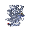
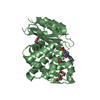
| Title | Crystal structure of the C-terminal deleted mutant of the class A carbapenemase KPC-2 at 1.23 angstrom | ||||||
 Components Components | Class A carbapenemase KPC-2 | ||||||
 Keywords Keywords | HYDROLASE / beta-lactamase / carbapenemase / C-terminal deleted mutant / Plasmid | ||||||
| Function / homology |  Function and homology information Function and homology informationbeta-lactam antibiotic catabolic process / beta-lactamase activity / beta-lactamase / response to antibiotic Similarity search - Function | ||||||
| Biological species |  | ||||||
| Method |  X-RAY DIFFRACTION / SYNCHROTRON / MOLECULAR REPLACEMENT / Resolution: 1.23 Å X-RAY DIFFRACTION / SYNCHROTRON / MOLECULAR REPLACEMENT / Resolution: 1.23 Å | ||||||
 Authors Authors | Petrella, S. / Ziental-Gelus, N. / Mayer, C. / Jarlier, V. / Sougakoff, W. | ||||||
 Citation Citation | Journal: ANTIMICROB.AGENTS CHEMOTHER. / Year: 2008 Title: Genetic and structural insights into the dissemination potential of the extremely-broad-spectrum class A {beta}-lactamase (EBSBL) KPC-2 identified in two strains of Escherichia coli and ...Title: Genetic and structural insights into the dissemination potential of the extremely-broad-spectrum class A {beta}-lactamase (EBSBL) KPC-2 identified in two strains of Escherichia coli and Enterobacter cloacae isolated from the same patient in France Authors: Petrella, S. / Ziental-Gelus, N. / Mayer, C. / Renard, M. / Jarlier, V. / Sougakoff, W. | ||||||
| History |
|
- Structure visualization
| Structure viewer | Molecule: MolmilJmol/JSmol |
|---|
- Downloads & links
-Download
| PDBx/mmCIF format | 3c5a.cif.gz | 114.7 KB | Display | PDBx/mmCIF format |
|---|---|---|---|---|
| PDB format | pdb3c5a.ent.gz | 86.9 KB | Display | PDB format |
| PDBx/mmJSON format | 3c5a.json.gz | Tree view | PDBx/mmJSON format | |
| Others |  Other downloads Other downloads |
-Validation report
| Summary document | 3c5a_validation.pdf.gz | 443.8 KB | Display | wwPDB validaton report |
|---|---|---|---|---|
| Full document | 3c5a_full_validation.pdf.gz | 445.2 KB | Display | |
| Data in XML | 3c5a_validation.xml.gz | 13.2 KB | Display | |
| Data in CIF | 3c5a_validation.cif.gz | 19.1 KB | Display | |
| Arichive directory | https://data.pdbj.org/pub/pdb/validation_reports/c5/3c5aftp://data.pdbj.org/pub/pdb/validation_reports/c5/3c5a | HTTPS FTP |
-Related structure data
| Related structure data |  2ods S: Starting model for refinement |
|---|---|
| Similar structure data |


-Links
 PDBj
PDBj


- Assembly
| Deposited unit | 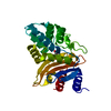
| ||||||||
|---|---|---|---|---|---|---|---|---|---|
| 1 |
| ||||||||
| Unit cell |
|
-Components
| #1: Protein | Mass: 27992.506 Da / Num. of mol.: 1 / Fragment: UNP residues 26-289 Source method: isolated from a genetically manipulated source Source: (gene. exp.) |
|---|---|
| #2: Chemical | ChemComp-CIT /   Mass: 192.124 Da / Num. of mol.: 1 / Source method: obtained synthetically / Formula: C6H8O7 Mass: 192.124 Da / Num. of mol.: 1 / Source method: obtained synthetically / Formula: C6H8O7 |
| #3: Water | ChemComp-HOH /  Mass: 18.015 Da / Num. of mol.: 183 / Source method: isolated from a natural source / Formula: H2O Mass: 18.015 Da / Num. of mol.: 183 / Source method: isolated from a natural source / Formula: H2O |
| Has protein modification | Y |
-Experimental details
-Experiment
| Experiment | Method: X-RAY DIFFRACTION / Number of used crystals: 1 |
|---|
- Sample preparation
| Crystal | Density Matthews: 2.04 Å3/Da / Density % sol: 39.58 % |
|---|---|
| Crystal grow | Temperature: 291 K / Method: vapor diffusion, sitting drop / pH: 4 Details: 20% PEG4K, 0.1M KSCN, 0.1M Citrate, VAPOR DIFFUSION, SITTING DROP, pH4.0, temperature 291K |
-Data collection
| Diffraction | Mean temperature: 77 K |
|---|---|
| Diffraction source | Source: SYNCHROTRON / Site: ESRF  / Beamline: BM30A / Wavelength: 0.979 Å / Beamline: BM30A / Wavelength: 0.979 Å |
| Detector | Type: ADSC QUANTUM 315 / Detector: CCD / Date: Mar 23, 2007 / Details: mirror 1, double crystal, mirror 2 |
| Radiation | Monochromator: Si 111 CHANNEL / Protocol: SINGLE WAVELENGTH / Monochromatic (M) / Laue (L): M / Scattering type: x-ray |
| Radiation wavelength | Wavelength: 0.979 Å / Relative weight: 1 |
| Reflection | Resolution: 1.23→27.17 Å / Num. obs: 65197 / % possible obs: 97.7 % / Redundancy: 13.6 % / Biso Wilson estimate: 14.7 Å2 / Net I/σ(I): 22.55 |
| Reflection shell | Resolution: 1.23→1.31 Å / Redundancy: 11.6 % / Mean I/σ(I) obs: 6.5 / Num. unique all: 9747 / Rsym value: 0.196 / % possible all: 91.9 |
- Processing
| Software |
| ||||||||||||||||||||||||||||||||||||||||||||||||||||||||||||||||||||||||||||||||||||||||||
|---|---|---|---|---|---|---|---|---|---|---|---|---|---|---|---|---|---|---|---|---|---|---|---|---|---|---|---|---|---|---|---|---|---|---|---|---|---|---|---|---|---|---|---|---|---|---|---|---|---|---|---|---|---|---|---|---|---|---|---|---|---|---|---|---|---|---|---|---|---|---|---|---|---|---|---|---|---|---|---|---|---|---|---|---|---|---|---|---|---|---|---|
| Refinement | Method to determine structure: MOLECULAR REPLACEMENT Starting model: PDB ENTRY 2ODS 2ods Resolution: 1.23→27.17 Å / Cor.coef. Fo:Fc: 0.959 / Cor.coef. Fo:Fc free: 0.954 / SU B: 0.644 / SU ML: 0.029 / Isotropic thermal model: RESTRAINED / Cross valid method: THROUGHOUT / σ(F): 0 / ESU R: 0.046 / ESU R Free: 0.046 / Stereochemistry target values: MAXIMUM LIKELIHOOD / Details: HYDROGENS HAVE BEEN ADDED IN THE RIDING POSITIONS
| ||||||||||||||||||||||||||||||||||||||||||||||||||||||||||||||||||||||||||||||||||||||||||
| Solvent computation | Ion probe radii: 0.8 Å / Shrinkage radii: 0.8 Å / VDW probe radii: 1.2 Å / Solvent model: MASK | ||||||||||||||||||||||||||||||||||||||||||||||||||||||||||||||||||||||||||||||||||||||||||
| Displacement parameters | Biso mean: 11.796 Å2
| ||||||||||||||||||||||||||||||||||||||||||||||||||||||||||||||||||||||||||||||||||||||||||
| Refinement step | Cycle: LAST / Resolution: 1.23→27.17 Å
| ||||||||||||||||||||||||||||||||||||||||||||||||||||||||||||||||||||||||||||||||||||||||||
| Refine LS restraints |
| ||||||||||||||||||||||||||||||||||||||||||||||||||||||||||||||||||||||||||||||||||||||||||
| LS refinement shell | Resolution: 1.233→1.265 Å / Total num. of bins used: 20
|
