 Movie
Movie Controller

[English] 日本語
 Yorodumi
- PDB-3c51: Crystal structure of G protein coupled receptor kinase 1 bound to... -
+ Open data

- Basic information
| Entry | Database: PDB / ID: 3c51 | ||||||
|---|---|---|---|---|---|---|---|
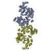
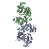
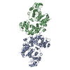
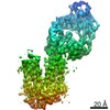
| Title | Crystal structure of G protein coupled receptor kinase 1 bound to ADP and magnesium chloride at 3.55A | ||||||
 Components Components | Rhodopsin kinase | ||||||
 Keywords Keywords | TRANSFERASE / Ser/Thr kinase / RGS homology domain / G protein coupled receptor kinase / GRK / GRK1 / rhodopsin kinase / P-loop / autophosphorylation / ADP / ATP-binding / Lipoprotein / Membrane / Methylation / Nucleotide-binding / Phosphoprotein / Prenylation / Serine/threonine-protein kinase | ||||||
| Function / homology |  Function and homology information Function and homology informationrhodopsin kinase / rhodopsin kinase activity / regulation of opsin-mediated signaling pathway / Inactivation, recovery and regulation of the phototransduction cascade / regulation of signal transduction / visual perception / photoreceptor disc membrane / protein autophosphorylation / signal transduction / ATP binding / cytoplasm Similarity search - Function | ||||||
| Biological species |  | ||||||
| Method |  X-RAY DIFFRACTION / SYNCHROTRON / MOLECULAR REPLACEMENT / Resolution: 3.55 Å X-RAY DIFFRACTION / SYNCHROTRON / MOLECULAR REPLACEMENT / Resolution: 3.55 Å | ||||||
 Authors Authors | Singh, P. / Tesmer, J.J.G. | ||||||
 Citation Citation | Journal: J.Biol.Chem. / Year: 2008 Title: Structures of rhodopsin kinase in different ligand states reveal key elements involved in G protein-coupled receptor kinase activation. Authors: Singh, P. / Wang, B. / Maeda, T. / Palczewski, K. / Tesmer, J.J. | ||||||
| History |
|
- Structure visualization
| Structure viewer | Molecule: MolmilJmol/JSmol |
|---|
- Downloads & links
-Download
| PDBx/mmCIF format | 3c51.cif.gz | 199.9 KB | Display | PDBx/mmCIF format |
|---|---|---|---|---|
| PDB format | pdb3c51.ent.gz | 157.4 KB | Display | PDB format |
| PDBx/mmJSON format | 3c51.json.gz | Tree view | PDBx/mmJSON format | |
| Others |  Other downloads Other downloads |
-Validation report
| Arichive directory | https://data.pdbj.org/pub/pdb/validation_reports/c5/3c51ftp://data.pdbj.org/pub/pdb/validation_reports/c5/3c51 | HTTPS FTP |
|---|
-Related structure data
| Related structure data | 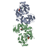 3c4wC  3c4xC  3c4yC  3c4zSC  3c50C C: citing same article ( S: Starting model for refinement |
|---|---|
| Similar structure data |


-Links
 PDBj
PDBj


- Assembly
| Deposited unit | 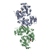
| |||||||||||||||||||||||||||||||||||||||||||||||||||||||||||||||||||||||||||||||||||||||||||
|---|---|---|---|---|---|---|---|---|---|---|---|---|---|---|---|---|---|---|---|---|---|---|---|---|---|---|---|---|---|---|---|---|---|---|---|---|---|---|---|---|---|---|---|---|---|---|---|---|---|---|---|---|---|---|---|---|---|---|---|---|---|---|---|---|---|---|---|---|---|---|---|---|---|---|---|---|---|---|---|---|---|---|---|---|---|---|---|---|---|---|---|---|
| 1 |
| |||||||||||||||||||||||||||||||||||||||||||||||||||||||||||||||||||||||||||||||||||||||||||
| Unit cell |
| |||||||||||||||||||||||||||||||||||||||||||||||||||||||||||||||||||||||||||||||||||||||||||
| Noncrystallographic symmetry (NCS) | NCS domain:
NCS domain segments: Ens-ID: 1 / Refine code: 3
|
-Components
| #1: Protein | Mass: 61429.934 Da / Num. of mol.: 2 / Fragment: UNP residues 1-535 Source method: isolated from a genetically manipulated source Source: (gene. exp.)  Trichoplusia ni (cabbage looper) / References: UniProt: P28327, rhodopsin kinase Trichoplusia ni (cabbage looper) / References: UniProt: P28327, rhodopsin kinase#2: Chemical | ChemComp-MG /   Mass: 24.305 Da / Num. of mol.: 4 / Source method: obtained synthetically / Formula: Mg Mass: 24.305 Da / Num. of mol.: 4 / Source method: obtained synthetically / Formula: Mg#3: Chemical | ChemComp-BR /   Mass: 79.904 Da / Num. of mol.: 5 / Source method: obtained synthetically / Formula: Br Mass: 79.904 Da / Num. of mol.: 5 / Source method: obtained synthetically / Formula: Br#4: Chemical |   Mass: 427.201 Da / Num. of mol.: 2 / Source method: obtained synthetically / Formula: C10H15N5O10P2 / Comment: ADP, energy-carrying molecule*YM Mass: 427.201 Da / Num. of mol.: 2 / Source method: obtained synthetically / Formula: C10H15N5O10P2 / Comment: ADP, energy-carrying molecule*YM |
|---|
-Experimental details
-Experiment
| Experiment | Method: X-RAY DIFFRACTION / Number of used crystals: 1 |
|---|
- Sample preparation
| Crystal | Density Matthews: 2.86 Å3/Da / Density % sol: 57.06 % |
|---|---|
| Crystal grow | Temperature: 277 K / Method: vapor diffusion, hanging drop / pH: 6.25 Details: PEG 8000, NaBr, glycerol, MES pH 6.25, ADP pH 7.5, magnesium chloride, VAPOR DIFFUSION, HANGING DROP, temperature 277K |
-Data collection
| Diffraction | Mean temperature: 100 K |
|---|---|
| Diffraction source | Source: SYNCHROTRON / Site: APS  / Beamline: 23-ID-D / Wavelength: 0.97934 Å / Beamline: 23-ID-D / Wavelength: 0.97934 Å |
| Detector | Type: MARMOSAIC 300 mm CCD / Detector: CCD / Date: Aug 17, 2006 |
| Radiation | Protocol: SINGLE WAVELENGTH / Monochromatic (M) / Laue (L): M / Scattering type: x-ray |
| Radiation wavelength | Wavelength: 0.97934 Å / Relative weight: 1 |
| Reflection | Resolution: 3.55→30 Å / Num. obs: 17511 |
- Processing
| Software |
| ||||||||||||||||||||||||||||||||||||||||||||||||||||||||||||||||||||||||||||||||||||||||||||||||||||||||||||||||||||||||||||||||||||||||||||||||||||||||||||||||||||||||||
|---|---|---|---|---|---|---|---|---|---|---|---|---|---|---|---|---|---|---|---|---|---|---|---|---|---|---|---|---|---|---|---|---|---|---|---|---|---|---|---|---|---|---|---|---|---|---|---|---|---|---|---|---|---|---|---|---|---|---|---|---|---|---|---|---|---|---|---|---|---|---|---|---|---|---|---|---|---|---|---|---|---|---|---|---|---|---|---|---|---|---|---|---|---|---|---|---|---|---|---|---|---|---|---|---|---|---|---|---|---|---|---|---|---|---|---|---|---|---|---|---|---|---|---|---|---|---|---|---|---|---|---|---|---|---|---|---|---|---|---|---|---|---|---|---|---|---|---|---|---|---|---|---|---|---|---|---|---|---|---|---|---|---|---|---|---|---|---|---|---|---|---|
| Refinement | Method to determine structure: MOLECULAR REPLACEMENT Starting model: PDB ENTRY 3C4Z Resolution: 3.55→19.88 Å / Cor.coef. Fo:Fc: 0.885 / SU B: 66.976 / SU ML: 0.509 / TLS residual ADP flag: LIKELY RESIDUAL / Stereochemistry target values: MAXIMUM LIKELIHOOD / Details: HYDROGENS HAVE BEEN ADDED IN THE RIDING POSITIONS
| ||||||||||||||||||||||||||||||||||||||||||||||||||||||||||||||||||||||||||||||||||||||||||||||||||||||||||||||||||||||||||||||||||||||||||||||||||||||||||||||||||||||||||
| Solvent computation | Ion probe radii: 0.8 Å / Shrinkage radii: 0.8 Å / VDW probe radii: 1.4 Å / Solvent model: MASK | ||||||||||||||||||||||||||||||||||||||||||||||||||||||||||||||||||||||||||||||||||||||||||||||||||||||||||||||||||||||||||||||||||||||||||||||||||||||||||||||||||||||||||
| Displacement parameters | Biso mean: 115.622 Å2
| ||||||||||||||||||||||||||||||||||||||||||||||||||||||||||||||||||||||||||||||||||||||||||||||||||||||||||||||||||||||||||||||||||||||||||||||||||||||||||||||||||||||||||
| Refinement step | Cycle: LAST / Resolution: 3.55→19.88 Å
| ||||||||||||||||||||||||||||||||||||||||||||||||||||||||||||||||||||||||||||||||||||||||||||||||||||||||||||||||||||||||||||||||||||||||||||||||||||||||||||||||||||||||||
| Refine LS restraints |
| ||||||||||||||||||||||||||||||||||||||||||||||||||||||||||||||||||||||||||||||||||||||||||||||||||||||||||||||||||||||||||||||||||||||||||||||||||||||||||||||||||||||||||
| Refine LS restraints NCS | Dom-ID: 1 / Auth asym-ID: A / Ens-ID: 1 / Refine-ID: X-RAY DIFFRACTION
| ||||||||||||||||||||||||||||||||||||||||||||||||||||||||||||||||||||||||||||||||||||||||||||||||||||||||||||||||||||||||||||||||||||||||||||||||||||||||||||||||||||||||||
| LS refinement shell | Resolution: 3.55→3.639 Å / Total num. of bins used: 20
| ||||||||||||||||||||||||||||||||||||||||||||||||||||||||||||||||||||||||||||||||||||||||||||||||||||||||||||||||||||||||||||||||||||||||||||||||||||||||||||||||||||||||||
| Refinement TLS params. | Method: refined / Refine-ID: X-RAY DIFFRACTION
| ||||||||||||||||||||||||||||||||||||||||||||||||||||||||||||||||||||||||||||||||||||||||||||||||||||||||||||||||||||||||||||||||||||||||||||||||||||||||||||||||||||||||||
| Refinement TLS group |
|
