 Movie
Movie Controller

[English] 日本語
 Yorodumi
- PDB-3c4s: Crystal structure of the Ssl0352 protein from Synechocystis sp. N... -
+ Open data

- Basic information
| Entry | Database: PDB / ID: 3c4s | ||||||
|---|---|---|---|---|---|---|---|
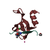
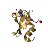
| Title | Crystal structure of the Ssl0352 protein from Synechocystis sp. Northeast Structural Genomics Consortium target SgR42 | ||||||
 Components Components | Ssl0352 protein | ||||||
 Keywords Keywords | STRUCTURAL GENOMICS / UNKNOWN FUNCTION / P74795_SYNY3 / Ssl0352 / NESG / SgR42 / PSI-2 / Protein Structure Initiative / Northeast Structural Genomics Consortium | ||||||
| Function / homology | NADH dehydrogenase-like complex, subunit S / NAD(P)H dehydrogenase subunit S / photosynthetic electron transport chain / SH3 type barrels. - #140 / SH3 type barrels. / Roll / Mainly Beta / Ssl0352 protein Function and homology information Function and homology information | ||||||
| Biological species |  | ||||||
| Method |  X-RAY DIFFRACTION / SYNCHROTRON / MOLECULAR REPLACEMENT / Resolution: 1.7 Å X-RAY DIFFRACTION / SYNCHROTRON / MOLECULAR REPLACEMENT / Resolution: 1.7 Å | ||||||
 Authors Authors | Vorobiev, S.M. / Chen, Y. / Seetharaman, J. / Wang, D. / Maglaqui, M. / Janjua, H. / Xiao, R. / Acton, T.B. / Montelione, G.T. / Hunt, J.F. ...Vorobiev, S.M. / Chen, Y. / Seetharaman, J. / Wang, D. / Maglaqui, M. / Janjua, H. / Xiao, R. / Acton, T.B. / Montelione, G.T. / Hunt, J.F. / Tong, L. / Northeast Structural Genomics Consortium (NESG) | ||||||
 Citation Citation | Journal: To be Published Title: Crystal structure of the Ssl0352 protein from Synechocystis sp. Authors: Vorobiev, S.M. / Chen, Y. / Seetharaman, J. / Wang, D. / Maglaqui, M. / Janjua, H. / Xiao, R. / Acton, T.B. / Montelione, G.T. / Hunt, J.F. / Tong, L. | ||||||
| History |
|
- Structure visualization
| Structure viewer | Molecule: MolmilJmol/JSmol |
|---|
- Downloads & links
-Download
| PDBx/mmCIF format | 3c4s.cif.gz | 38.4 KB | Display | PDBx/mmCIF format |
|---|---|---|---|---|
| PDB format | pdb3c4s.ent.gz | 25.5 KB | Display | PDB format |
| PDBx/mmJSON format | 3c4s.json.gz | Tree view | PDBx/mmJSON format | |
| Others |  Other downloads Other downloads |
-Validation report
| Arichive directory | https://data.pdbj.org/pub/pdb/validation_reports/c4/3c4sftp://data.pdbj.org/pub/pdb/validation_reports/c4/3c4s | HTTPS FTP |
|---|
-Related structure data
| Related structure data |  2jz2S S: Starting model for refinement |
|---|---|
| Similar structure data | |
| Other databases |


-Links
 PDBj
PDBj- Assembly
| Deposited unit | 
| ||||||||
|---|---|---|---|---|---|---|---|---|---|
| 1 | 
| ||||||||
| 2 | 
| ||||||||
| Unit cell |
| ||||||||
| Details | AUTHORS STATE THAT THE BIOLOGICAL UNIT IS A MONOMER ACCORDING TO AGGREGATION SCREENING. |
-Components
| #1: Protein | Mass: 7655.642 Da / Num. of mol.: 2 Source method: isolated from a genetically manipulated source Source: (gene. exp.) #2: Water | ChemComp-HOH / |  Mass: 18.015 Da / Num. of mol.: 149 / Source method: isolated from a natural source / Formula: H2O Mass: 18.015 Da / Num. of mol.: 149 / Source method: isolated from a natural source / Formula: H2O |
|---|
-Experimental details
-Experiment
| Experiment | Method: X-RAY DIFFRACTION / Number of used crystals: 1 |
|---|
- Sample preparation
| Crystal | Density Matthews: 2.44 Å3/Da / Density % sol: 49.63 % Description: The structure factor file contains Friedel pairs |
|---|---|
| Crystal grow | Temperature: 291 K / Method: microbatch under oil / pH: 6.5 Details: 30% Jeffamine M-600, 0.05M Cesium chloride, 0.1M MES pH 6.5, MICROBATCH UNDER OIL, temperature 291K |
-Data collection
| Diffraction | Mean temperature: 100 K |
|---|---|
| Diffraction source | Source: SYNCHROTRON / Site: NSLS  / Beamline: X4C / Wavelength: 0.979 Å / Beamline: X4C / Wavelength: 0.979 Å |
| Detector | Type: MAR scanner 345 mm plate / Detector: IMAGE PLATE / Date: Jan 25, 2008 |
| Radiation | Protocol: SINGLE WAVELENGTH / Monochromatic (M) / Laue (L): M / Scattering type: x-ray |
| Radiation wavelength | Wavelength: 0.979 Å / Relative weight: 1 |
| Reflection | Resolution: 1.7→50 Å / Num. all: 31765 / Num. obs: 31765 / % possible obs: 99.3 % / Observed criterion σ(F): 0 / Observed criterion σ(I): 0 / Redundancy: 3.9 % / Biso Wilson estimate: 14.1 Å2 / Rmerge(I) obs: 0.057 / Net I/σ(I): 17.36 |
| Reflection shell | Resolution: 1.7→1.76 Å / Redundancy: 3.8 % / Rmerge(I) obs: 0.145 / Mean I/σ(I) obs: 6 / Num. unique all: 3200 / % possible all: 100 |
- Processing
| Software |
| ||||||||||||||||||||||||||||||||||||||||||||||||||||||||||||
|---|---|---|---|---|---|---|---|---|---|---|---|---|---|---|---|---|---|---|---|---|---|---|---|---|---|---|---|---|---|---|---|---|---|---|---|---|---|---|---|---|---|---|---|---|---|---|---|---|---|---|---|---|---|---|---|---|---|---|---|---|---|
| Refinement | Method to determine structure: MOLECULAR REPLACEMENT Starting model: PDB entry 2JZ2, chain A Resolution: 1.7→24.89 Å / Rfactor Rfree error: 0.005 / Data cutoff high absF: 1019206.95 / Data cutoff low absF: 0 / Isotropic thermal model: RESTRAINED / Cross valid method: THROUGHOUT / σ(F): 2 / Stereochemistry target values: Engh & Huber / Details: The Friedel pairs were used in phasing
| ||||||||||||||||||||||||||||||||||||||||||||||||||||||||||||
| Solvent computation | Solvent model: FLAT MODEL / Bsol: 69.6645 Å2 / ksol: 0.421479 e/Å3 | ||||||||||||||||||||||||||||||||||||||||||||||||||||||||||||
| Displacement parameters | Biso mean: 20 Å2
| ||||||||||||||||||||||||||||||||||||||||||||||||||||||||||||
| Refine analyze |
| ||||||||||||||||||||||||||||||||||||||||||||||||||||||||||||
| Refinement step | Cycle: LAST / Resolution: 1.7→24.89 Å
| ||||||||||||||||||||||||||||||||||||||||||||||||||||||||||||
| Refine LS restraints |
| ||||||||||||||||||||||||||||||||||||||||||||||||||||||||||||
| LS refinement shell | Resolution: 1.7→1.81 Å / Rfactor Rfree error: 0.014 / Total num. of bins used: 6
| ||||||||||||||||||||||||||||||||||||||||||||||||||||||||||||
| Xplor file |
|
