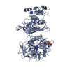
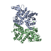
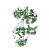
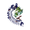
Entry Database : PDB / ID : 3c4bTitle Structure of RNaseIIIb and dsRNA binding domains of mouse Dicer Endoribonuclease Dicer Keywords / / / / / / / / / / Function / homology Function Domain/homology Component
/ / / / / / / / / / / / / / / / / / / / / / / / / / / / / / / / / / / / / / / / / / / / / / / / / / / / / / / / / / / / / / / / / / / / / / / / / / / / / / / / / / / / / / / / / / / / / / / / / / / / / / / / / / / / / / / / / / / / / / / / / / / / / / / / / / / / / / / / / / / / / / / / / / / / / / / / / / / / / / / Biological species Mus musculus (house mouse)Method / / Resolution : 1.68 Å Authors Lee, J.K. / Du, Z. / Tjhen, R.J. / Stroud, R.M. / James, T.L. Journal : Proc.Natl.Acad.Sci.Usa / Year : 2008Title : Structural and biochemical insights into the dicing mechanism of mouse Dicer: A conserved lysine is critical for dsRNA cleavage.Authors : Du, Z. / Lee, J.K. / Tjhen, R. / Stroud, R.M. / James, T.L. History Deposition Jan 29, 2008 Deposition site / Processing site Revision 1.0 Feb 19, 2008 Provider / Type Revision 1.1 Jul 13, 2011 Group / Version format complianceRevision 1.2 Mar 26, 2025 Group Data collection / Database references ... Data collection / Database references / Derived calculations / Structure summary Category chem_comp_atom / chem_comp_bond ... chem_comp_atom / chem_comp_bond / database_2 / pdbx_entry_details / pdbx_modification_feature / struct_conn / struct_ref_seq_dif Item _database_2.pdbx_DOI / _database_2.pdbx_database_accession ... _database_2.pdbx_DOI / _database_2.pdbx_database_accession / _struct_conn.pdbx_leaving_atom_flag / _struct_ref_seq_dif.details
Show all Show less
 Movie
Movie Controller


 Open data
 Basic information
 Components
 Keywords
 Function and homology information
 X-RAY DIFFRACTION /
  Authors
 Citation
 Structure visualization
 Downloads & links
 Other downloads


 PDBj
PDBj

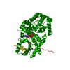
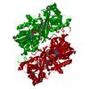
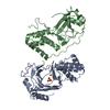
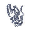
 Assembly


 Mass: 18.015 Da / Num. of mol.: 307 / Source method: isolated from a natural source / Formula: H2O
 Sample preparation
 Processing