 Movie
Movie Controller


+ Open data

- Basic information
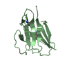
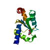
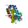
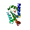
| Entry | Database: PDB / ID: 3c1s | ||||||
|---|---|---|---|---|---|---|---|
| Title | Crystal structure of GRX1 in glutathionylated form | ||||||
 Components Components | Glutaredoxin-1 | ||||||
 Keywords Keywords | OXIDOREDUCTASE / GLUTATHIONYLATED FORM | ||||||
| Function / homology |  Function and homology information Function and homology informationglutathione peroxidase / glutathione disulfide oxidoreductase activity / glutathione peroxidase activity / glutathione transferase / glutathione transferase activity / cellular response to oxidative stress / nucleus / cytoplasm Similarity search - Function | ||||||
| Biological species |  | ||||||
| Method |  X-RAY DIFFRACTION / MOLECULAR REPLACEMENT / Resolution: 2.5 Å X-RAY DIFFRACTION / MOLECULAR REPLACEMENT / Resolution: 2.5 Å | ||||||
 Authors Authors | Yu, J. / Zhou, C.Z. | ||||||
 Citation Citation | Journal: Proteins / Year: 2008 Title: Glutathionylation-triggered conformational changes of glutaredoxin Grx1 from the yeast Saccharomyces cerevisiae. Authors: Yu, J. / Zhang, N.N. / Yin, P.D. / Cui, P.X. / Zhou, C.Z. | ||||||
| History |
|
- Structure visualization
| Structure viewer | Molecule: MolmilJmol/JSmol |
|---|
- Downloads & links
-Download
| PDBx/mmCIF format | 3c1s.cif.gz | 36.7 KB | Display | PDBx/mmCIF format |
|---|---|---|---|---|
| PDB format | pdb3c1s.ent.gz | 23.5 KB | Display | PDB format |
| PDBx/mmJSON format | 3c1s.json.gz | Tree view | PDBx/mmJSON format | |
| Others |  Other downloads Other downloads |
-Validation report
| Arichive directory | https://data.pdbj.org/pub/pdb/validation_reports/c1/3c1sftp://data.pdbj.org/pub/pdb/validation_reports/c1/3c1s | HTTPS FTP |
|---|
-Related structure data
| Related structure data | 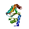 3c1rSC S: Starting model for refinement C: citing same article ( |
|---|---|
| Similar structure data |


-Links
 PDBj
PDBj


- Assembly
| Deposited unit | 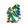
| ||||||||
|---|---|---|---|---|---|---|---|---|---|
| 1 |
| ||||||||
| Unit cell |
|
-Components
| #1: Protein | Mass: 13411.281 Da / Num. of mol.: 1 Source method: isolated from a genetically manipulated source Source: (gene. exp.) Strain: S288C / Gene: GRX1 / Plasmid: PET28B / Production host:  References: UniProt: P25373, arsenate reductase (glutathione/glutaredoxin) |
|---|---|
| #2: Chemical | ChemComp-GSH /   Mass: 307.323 Da / Num. of mol.: 1 / Source method: obtained synthetically / Formula: C10H17N3O6S Mass: 307.323 Da / Num. of mol.: 1 / Source method: obtained synthetically / Formula: C10H17N3O6S |
| #3: Water | ChemComp-HOH /  Mass: 18.015 Da / Num. of mol.: 73 / Source method: isolated from a natural source / Formula: H2O Mass: 18.015 Da / Num. of mol.: 73 / Source method: isolated from a natural source / Formula: H2O |
-Experimental details
-Experiment
| Experiment | Method: X-RAY DIFFRACTION / Number of used crystals: 1 |
|---|
- Sample preparation
| Crystal | Density Matthews: 2.33 Å3/Da / Density % sol: 47.29 % |
|---|---|
| Crystal grow | Temperature: 289 K / Method: vapor diffusion, hanging drop / pH: 5.3 Details: 20% PEG 4000, 0.1M SODIUM ACETATE, 0.2M AMMONIUM DIHYDROGEN PHOSPHATE, pH 5.30, VAPOR DIFFUSION, HANGING DROP, temperature 289K |
-Data collection
| Diffraction | Mean temperature: 100 K |
|---|---|
| Diffraction source | Source: ROTATING ANODE / Type: RIGAKU / Wavelength: 1.5418 Å |
| Detector | Type: MAR scanner 345 mm plate / Detector: IMAGE PLATE / Details: MIRRORS |
| Radiation | Monochromator: YALE MIRRORS / Protocol: SINGLE WAVELENGTH / Monochromatic (M) / Laue (L): M / Scattering type: x-ray |
| Radiation wavelength | Wavelength: 1.5418 Å / Relative weight: 1 |
| Reflection | Resolution: 2.5→29.74 Å / Num. obs: 4547 / % possible obs: 97.9 % / Observed criterion σ(I): 1 / Redundancy: 2.79 % / Biso Wilson estimate: 42.8 Å2 / Rmerge(I) obs: 0.0918 / Net I/σ(I): 4.3 |
| Reflection shell | Resolution: 2.5→2.59 Å / Redundancy: 2.82 % / Rmerge(I) obs: 0.1787 / Mean I/σ(I) obs: 2.9 / Num. unique all: 418 / % possible all: 99.8 |
- Processing
| Software |
| ||||||||||||||||||||||||||||||||||||||||||||||||||||||||||||
|---|---|---|---|---|---|---|---|---|---|---|---|---|---|---|---|---|---|---|---|---|---|---|---|---|---|---|---|---|---|---|---|---|---|---|---|---|---|---|---|---|---|---|---|---|---|---|---|---|---|---|---|---|---|---|---|---|---|---|---|---|---|
| Refinement | Method to determine structure: MOLECULAR REPLACEMENT Starting model: PDB ENTRY 3C1R Resolution: 2.5→29.74 Å / Rfactor Rfree error: 0.017 / Data cutoff high absF: 883820.03 / Data cutoff low absF: 0 / Isotropic thermal model: RESTRAINED / Cross valid method: THROUGHOUT / σ(F): 0 / Stereochemistry target values: Engh & Huber
| ||||||||||||||||||||||||||||||||||||||||||||||||||||||||||||
| Solvent computation | Solvent model: FLAT MODEL / Bsol: 41.92 Å2 / ksol: 0.36 e/Å3 | ||||||||||||||||||||||||||||||||||||||||||||||||||||||||||||
| Displacement parameters | Biso mean: 31.9 Å2
| ||||||||||||||||||||||||||||||||||||||||||||||||||||||||||||
| Refine analyze |
| ||||||||||||||||||||||||||||||||||||||||||||||||||||||||||||
| Refinement step | Cycle: LAST / Resolution: 2.5→29.74 Å
| ||||||||||||||||||||||||||||||||||||||||||||||||||||||||||||
| Refine LS restraints |
| ||||||||||||||||||||||||||||||||||||||||||||||||||||||||||||
| LS refinement shell | Resolution: 2.5→2.59 Å / Rfactor Rfree error: 0.075 / Total num. of bins used: 10
| ||||||||||||||||||||||||||||||||||||||||||||||||||||||||||||
| Xplor file |
|
