 Movie
Movie Controller

[English] 日本語
 Yorodumi
- PDB-3bv7: Crystal structure of Delta(4)-3-ketosteroid 5-beta-reductase in c... -
+ Open data

- Basic information
| Entry | Database: PDB / ID: 3bv7 | ||||||
|---|---|---|---|---|---|---|---|
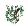
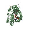
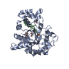
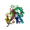
| Title | Crystal structure of Delta(4)-3-ketosteroid 5-beta-reductase in complex with NADP and glycerol. Resolution: 1.79 A. | ||||||
 Components Components | 3-oxo-5-beta-steroid 4-dehydrogenase | ||||||
 Keywords Keywords | OXIDOREDUCTASE / 5-beta-reductase / glycerol / AKR1A1 / Bile acid catabolism / Disease mutation / Lipid metabolism / NADP / Steroid metabolism | ||||||
| Function / homology |  Function and homology information Function and homology informationDelta4-3-oxosteroid 5beta-reductase / steroid dehydrogenase activity / bile acid catabolic process / C21-steroid hormone metabolic process / Delta4-3-oxosteroid 5beta-reductase activity / ketosteroid monooxygenase activity / Synthesis of bile acids and bile salts via 24-hydroxycholesterol / bile acid biosynthetic process / alcohol dehydrogenase (NADP+) activity / cholesterol catabolic process ...Delta4-3-oxosteroid 5beta-reductase / steroid dehydrogenase activity / bile acid catabolic process / C21-steroid hormone metabolic process / Delta4-3-oxosteroid 5beta-reductase activity / ketosteroid monooxygenase activity / Synthesis of bile acids and bile salts via 24-hydroxycholesterol / bile acid biosynthetic process / alcohol dehydrogenase (NADP+) activity / cholesterol catabolic process / aldose reductase (NADPH) activity / androgen metabolic process / Synthesis of bile acids and bile salts via 27-hydroxycholesterol / Synthesis of bile acids and bile salts via 7alpha-hydroxycholesterol / digestion / steroid binding / cytosol Similarity search - Function | ||||||
| Biological species |  Homo sapiens (human) Homo sapiens (human) | ||||||
| Method |  X-RAY DIFFRACTION / SYNCHROTRON / FOURIER SYNTHESIS / Resolution: 1.79 Å X-RAY DIFFRACTION / SYNCHROTRON / FOURIER SYNTHESIS / Resolution: 1.79 Å | ||||||
 Authors Authors | Di Costanzo, L. / Drury, J. / Penning, T.M. / Christianson, D.W. | ||||||
 Citation Citation | Journal: J.Biol.Chem. / Year: 2008 Title: Crystal Structure of Human Liver {Delta}4-3-Ketosteroid 5{beta}-Reductase (AKR1D1) and Implications for Substrate Binding and Catalysis. Authors: Di Costanzo, L. / Drury, J.E. / Penning, T.M. / Christianson, D.W. | ||||||
| History |
|
- Structure visualization
| Structure viewer | Molecule: MolmilJmol/JSmol |
|---|
- Downloads & links
-Download
| PDBx/mmCIF format | 3bv7.cif.gz | 151.7 KB | Display | PDBx/mmCIF format |
|---|---|---|---|---|
| PDB format | pdb3bv7.ent.gz | 118.5 KB | Display | PDB format |
| PDBx/mmJSON format | 3bv7.json.gz | Tree view | PDBx/mmJSON format | |
| Others |  Other downloads Other downloads |
-Validation report
| Arichive directory | https://data.pdbj.org/pub/pdb/validation_reports/bv/3bv7ftp://data.pdbj.org/pub/pdb/validation_reports/bv/3bv7 | HTTPS FTP |
|---|
-Related structure data
| Related structure data |  3burSC  3buvC  3cmfC  3cotC S: Starting model for refinement C: citing same article ( |
|---|---|
| Similar structure data |


-Links
 PDBj
PDBj


- Assembly
| Deposited unit | 
| ||||||||
|---|---|---|---|---|---|---|---|---|---|
| 1 | 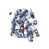
| ||||||||
| 2 | 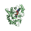
| ||||||||
| Unit cell |
|
-Components
| #1: Protein | Mass: 37427.898 Da / Num. of mol.: 2 Source method: isolated from a genetically manipulated source Source: (gene. exp.) Homo sapiens (human) / Gene: AKR1D1, SRD5B1 / Organ: liver / Plasmid: pET28a-AKR1D1 / Production host:  References: UniProt: P51857, Delta4-3-oxosteroid 5beta-reductase #2: Chemical |   Mass: 743.405 Da / Num. of mol.: 2 / Source method: obtained synthetically / Formula: C21H28N7O17P3 Mass: 743.405 Da / Num. of mol.: 2 / Source method: obtained synthetically / Formula: C21H28N7O17P3#3: Chemical |   Mass: 92.094 Da / Num. of mol.: 3 / Source method: obtained synthetically / Formula: C3H8O3 Mass: 92.094 Da / Num. of mol.: 3 / Source method: obtained synthetically / Formula: C3H8O3#4: Water | ChemComp-HOH / |  Mass: 18.015 Da / Num. of mol.: 348 / Source method: isolated from a natural source / Formula: H2O Mass: 18.015 Da / Num. of mol.: 348 / Source method: isolated from a natural source / Formula: H2O |
|---|
-Experimental details
-Experiment
| Experiment | Method: X-RAY DIFFRACTION / Number of used crystals: 1 |
|---|
- Sample preparation
| Crystal | Density Matthews: 2.36 Å3/Da / Density % sol: 47.91 % |
|---|---|
| Crystal grow | Temperature: 277 K / Method: vapor diffusion, hanging drop / pH: 7.5 Details: Protein: 5.0 mg/mL AKR1D1, 2.0 mM NADPH, 2.0 mM 5-beta-cholestan-3-one, 10.0 mM Tris (pH 7.4). Precipitant buffer: 0.1 M Tris (pH 7.0), 10-20% (wt/vol) PEG 4000, 10% isopropanol., pH 7.5, ...Details: Protein: 5.0 mg/mL AKR1D1, 2.0 mM NADPH, 2.0 mM 5-beta-cholestan-3-one, 10.0 mM Tris (pH 7.4). Precipitant buffer: 0.1 M Tris (pH 7.0), 10-20% (wt/vol) PEG 4000, 10% isopropanol., pH 7.5, VAPOR DIFFUSION, HANGING DROP, temperature 277K |
-Data collection
| Diffraction | Mean temperature: 100 K |
|---|---|
| Diffraction source | Source: SYNCHROTRON / Site: APS  / Beamline: 22-BM / Wavelength: 1 Å / Beamline: 22-BM / Wavelength: 1 Å |
| Detector | Type: MARMOSAIC 225 mm CCD / Detector: CCD / Date: Nov 23, 2007 Details: Rosenbaum-Rock monochromator high-resolution double-crystal Si (111) sagittal focusing. Rosenbaum-Rock vertical focusing mirror. |
| Radiation | Protocol: SINGLE WAVELENGTH / Monochromatic (M) / Laue (L): M / Scattering type: x-ray |
| Radiation wavelength | Wavelength: 1 Å / Relative weight: 1 |
| Reflection | Resolution: 1.79→50 Å / Num. all: 66774 / Num. obs: 66774 / % possible obs: 98.6 % / Redundancy: 4.1 % / Rmerge(I) obs: 0.109 / Net I/σ(I): 20.5 |
| Reflection shell | Resolution: 1.79→1.85 Å / Redundancy: 4.1 % / Rmerge(I) obs: 0.37 / Mean I/σ(I) obs: 3.4 / Num. unique all: 6684 / % possible all: 100 |
- Processing
| Software |
| |||||||||||||||||||||
|---|---|---|---|---|---|---|---|---|---|---|---|---|---|---|---|---|---|---|---|---|---|---|
| Refinement | Method to determine structure: FOURIER SYNTHESIS Starting model: 3BUR Resolution: 1.79→50 Å
| |||||||||||||||||||||
| Refinement step | Cycle: LAST / Resolution: 1.79→50 Å
| |||||||||||||||||||||
| Refine LS restraints |
|
