 Movie
Movie Controller

[English] 日本語
 Yorodumi
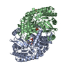
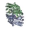
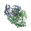
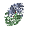
- PDB-3bv0: Crystal Structure of PLP Bound 7,8-Diaminopelargonic Acid Synthas... -
+ Open data

- Basic information
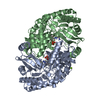
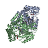
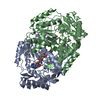
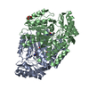
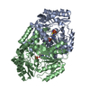
| Entry | Database: PDB / ID: 3bv0 | ||||||
|---|---|---|---|---|---|---|---|
| Title | Crystal Structure of PLP Bound 7,8-Diaminopelargonic Acid Synthase in Mycobacterium Tuberculosis | ||||||
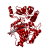
 Components Components | Adenosylmethionine-8-amino-7-oxononanoate aminotransferase | ||||||
 Keywords Keywords | TRANSFERASE / aminotransferase / biotin biosynthesis / Pyridoxal phosphate / S-adenosyl-L-methionine | ||||||
| Function / homology |  Function and homology information Function and homology informationadenosylmethionine-8-amino-7-oxononanoate transaminase / S-adenosyl-L-methionine:8-amino-7-oxononanoate transaminase activity / biotin biosynthetic process / pyridoxal phosphate binding / cytoplasm Similarity search - Function | ||||||
| Biological species |   Mycobacterium tuberculosis (bacteria) Mycobacterium tuberculosis (bacteria) | ||||||
| Method |  X-RAY DIFFRACTION / MOLECULAR REPLACEMENT / Resolution: 2.21 Å X-RAY DIFFRACTION / MOLECULAR REPLACEMENT / Resolution: 2.21 Å | ||||||
 Authors Authors | Dey, S. / Sacchettini, J.C. | ||||||
 Citation Citation | Journal: Biochemistry / Year: 2010 Title: Structural characterization of the Mycobacterium tuberculosis biotin biosynthesis enzymes 7,8-diaminopelargonic acid synthase and dethiobiotin synthetase . Authors: Dey, S. / Lane, J.M. / Lee, R.E. / Rubin, E.J. / Sacchettini, J.C. | ||||||
| History |
|
- Structure visualization
| Structure viewer | Molecule: MolmilJmol/JSmol |
|---|
- Downloads & links
-Download
| PDBx/mmCIF format | 3bv0.cif.gz | 168.1 KB | Display | PDBx/mmCIF format |
|---|---|---|---|---|
| PDB format | pdb3bv0.ent.gz | 132.4 KB | Display | PDB format |
| PDBx/mmJSON format | 3bv0.json.gz | Tree view | PDBx/mmJSON format | |
| Others |  Other downloads Other downloads |
-Validation report
| Arichive directory | https://data.pdbj.org/pub/pdb/validation_reports/bv/3bv0ftp://data.pdbj.org/pub/pdb/validation_reports/bv/3bv0 | HTTPS FTP |
|---|
-Related structure data
| Related structure data |  3dodC  3drdC  3du4C 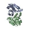 3fgnC 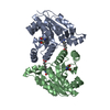 3fmfC 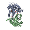 3fmiC 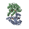 3fpaC  3lv2C  1qj5S S: Starting model for refinement C: citing same article ( |
|---|---|
| Similar structure data |
-Links
 PDBj
PDBj- Assembly
| Deposited unit | 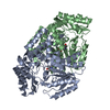
| ||||||||
|---|---|---|---|---|---|---|---|---|---|
| 1 |
| ||||||||
| Unit cell |
|
-Components
| #1: Protein | Mass: 46384.215 Da / Num. of mol.: 2 / Mutation: H315R Source method: isolated from a genetically manipulated source Details: DATABASE REFERENCE: TUBERCULIST / Source: (gene. exp.) Mycobacterium tuberculosis (bacteria) / Gene: bioA, Rv1568 / Plasmid: pET28b / Production host: References: UniProt: P0A4X6, UniProt: P9WQ81*PLUS, adenosylmethionine-8-amino-7-oxononanoate transaminase #2: Chemical |   Mass: 247.142 Da / Num. of mol.: 2 / Source method: obtained synthetically / Formula: C8H10NO6P Mass: 247.142 Da / Num. of mol.: 2 / Source method: obtained synthetically / Formula: C8H10NO6P#3: Water | ChemComp-HOH / |  Mass: 18.015 Da / Num. of mol.: 245 / Source method: isolated from a natural source / Formula: H2O Mass: 18.015 Da / Num. of mol.: 245 / Source method: isolated from a natural source / Formula: H2OSequence details | AUTHORS STATE THAT H315R WAS A NATURAL MUTATION | |
|---|
-Experimental details
-Experiment
| Experiment | Method: X-RAY DIFFRACTION / Number of used crystals: 1 |
|---|
- Sample preparation
| Crystal | Density Matthews: 2.3 Å3/Da / Density % sol: 46.47 % |
|---|---|
| Crystal grow | Temperature: 291 K / Method: vapor diffusion / pH: 7 Details: PEG 8000, 0.1M Tris buffer, 0.1M MgCl2, pH 7.0, VAPOR DIFFUSION, temperature 291K |
-Data collection
| Diffraction | Mean temperature: 100 K |
|---|---|
| Diffraction source | Source: ROTATING ANODE / Type: OTHER / Wavelength: 1.541 Å |
| Detector | Type: RIGAKU RAXIS IV++ / Detector: IMAGE PLATE |
| Radiation | Protocol: SINGLE WAVELENGTH / Monochromatic (M) / Laue (L): M / Scattering type: x-ray |
| Radiation wavelength | Wavelength: 1.541 Å / Relative weight: 1 |
| Reflection | Resolution: 2.21→35 Å / Num. obs: 40606 / % possible obs: 92.9 % / Redundancy: 6.2 % / Rmerge(I) obs: 0.073 / Net I/σ(I): 21.3 |
| Reflection shell | Resolution: 2.21→2.33 Å / Redundancy: 3.6 % / Rmerge(I) obs: 0.31 / Mean I/σ(I) obs: 3.5 / % possible all: 78.4 |
- Processing
| Software |
| ||||||||||||||||||||||||||||||||||||||||||||||||||||||||||||||||||||||||||||||||||||||||||||||||||||||||||||||||||||||||||||||||||||||||||||||||||||||||||||||||||||||||||
|---|---|---|---|---|---|---|---|---|---|---|---|---|---|---|---|---|---|---|---|---|---|---|---|---|---|---|---|---|---|---|---|---|---|---|---|---|---|---|---|---|---|---|---|---|---|---|---|---|---|---|---|---|---|---|---|---|---|---|---|---|---|---|---|---|---|---|---|---|---|---|---|---|---|---|---|---|---|---|---|---|---|---|---|---|---|---|---|---|---|---|---|---|---|---|---|---|---|---|---|---|---|---|---|---|---|---|---|---|---|---|---|---|---|---|---|---|---|---|---|---|---|---|---|---|---|---|---|---|---|---|---|---|---|---|---|---|---|---|---|---|---|---|---|---|---|---|---|---|---|---|---|---|---|---|---|---|---|---|---|---|---|---|---|---|---|---|---|---|---|---|---|
| Refinement | Method to determine structure: MOLECULAR REPLACEMENT Starting model: PDB entry 1QJ5 Resolution: 2.21→35 Å / Cor.coef. Fo:Fc: 0.939 / Cor.coef. Fo:Fc free: 0.901 / SU B: 5.304 / SU ML: 0.139 / Cross valid method: THROUGHOUT / ESU R: 0.308 / ESU R Free: 0.225 / Stereochemistry target values: MAXIMUM LIKELIHOOD
| ||||||||||||||||||||||||||||||||||||||||||||||||||||||||||||||||||||||||||||||||||||||||||||||||||||||||||||||||||||||||||||||||||||||||||||||||||||||||||||||||||||||||||
| Solvent computation | Ion probe radii: 0.8 Å / Shrinkage radii: 0.8 Å / VDW probe radii: 1.4 Å / Solvent model: MASK | ||||||||||||||||||||||||||||||||||||||||||||||||||||||||||||||||||||||||||||||||||||||||||||||||||||||||||||||||||||||||||||||||||||||||||||||||||||||||||||||||||||||||||
| Displacement parameters | Biso mean: 22.445 Å2
| ||||||||||||||||||||||||||||||||||||||||||||||||||||||||||||||||||||||||||||||||||||||||||||||||||||||||||||||||||||||||||||||||||||||||||||||||||||||||||||||||||||||||||
| Refinement step | Cycle: LAST / Resolution: 2.21→35 Å
| ||||||||||||||||||||||||||||||||||||||||||||||||||||||||||||||||||||||||||||||||||||||||||||||||||||||||||||||||||||||||||||||||||||||||||||||||||||||||||||||||||||||||||
| Refine LS restraints |
| ||||||||||||||||||||||||||||||||||||||||||||||||||||||||||||||||||||||||||||||||||||||||||||||||||||||||||||||||||||||||||||||||||||||||||||||||||||||||||||||||||||||||||
| LS refinement shell | Resolution: 2.21→2.267 Å / Total num. of bins used: 20
|
