- PDB-3bpd: Crystal structure of an uncharacterized protein (O28723_ARCFU) fr... -
+
Open data
ID or keywords:
Loading...
-
Basic information
Entry
Database: PDB / ID: 3bpd
Title
Crystal structure of an uncharacterized protein (O28723_ARCFU) from Archaeoglobus fulgidus
Components
Uncharacterized protein
Keywords
STRUCTURAL GENOMICS / UNKNOWN FUNCTION / heptamer / Mg+2 ion / 10197b / PSI-2 / NYSGXRC / Protein Structure Initiative / New York SGX Research Center for Structural Genomics
Function / homology
MTH889-like domain / Protein of unknown function DUF211 / MTH889-like domain superfamily / Uncharacterized ArCR, COG1888 / Alpha-Beta Plaits / 2-Layer Sandwich / metal ion binding / Alpha Beta / DUF211 domain-containing protein
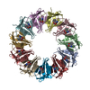
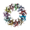
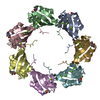
A: Uncharacterized protein B: Uncharacterized protein C: Uncharacterized protein D: Uncharacterized protein E: Uncharacterized protein F: Uncharacterized protein G: Uncharacterized protein H: Uncharacterized protein I: Uncharacterized protein J: Uncharacterized protein K: Uncharacterized protein L: Uncharacterized protein M: Uncharacterized protein N: Uncharacterized protein hetero molecules
A: Uncharacterized protein B: Uncharacterized protein C: Uncharacterized protein D: Uncharacterized protein E: Uncharacterized protein F: Uncharacterized protein G: Uncharacterized protein hetero molecules
H: Uncharacterized protein I: Uncharacterized protein J: Uncharacterized protein K: Uncharacterized protein L: Uncharacterized protein M: Uncharacterized protein N: Uncharacterized protein hetero molecules
Resolution: 2.8→50 Å / Cross valid method: THROUGHOUT / σ(F): 0 / Stereochemistry target values: Engh & Huber Details: The Friedel pairs were used in phasing. Residues listed as missing in Remark 465 are due to lack of electron density. Residues with missing atoms listed in Remark 470 are due to lack of ...Details: The Friedel pairs were used in phasing. Residues listed as missing in Remark 465 are due to lack of electron density. Residues with missing atoms listed in Remark 470 are due to lack of electron density for side chains and modeled as alanines.
Rfactor
Num. reflection
% reflection
Selection details
Rfree
0.305
1940
-
RANDOM
Rwork
0.242
-
-
-
all
-
38563
-
-
obs
-
38563
99.3 %
-
Displacement parameters
Biso mean: 36.4 Å2
Baniso -1
Baniso -2
Baniso -3
1-
1.74 Å2
0 Å2
0 Å2
2-
-
-0.8 Å2
0 Å2
3-
-
-
-0.93 Å2
Refine analyze
Free
Obs
Luzzati coordinate error
0.58 Å
0.42 Å
Luzzati d res low
-
5 Å
Luzzati sigma a
0.6 Å
0.45 Å
Refinement step
Cycle: LAST / Resolution: 2.8→50 Å
Protein
Nucleic acid
Ligand
Solvent
Total
Num. atoms
9804
0
27
91
9922
Refine LS restraints
Refine-ID
Type
Dev ideal
X-RAY DIFFRACTION
c_bond_d
0.008
X-RAY DIFFRACTION
c_angle_deg
1.7
X-RAY DIFFRACTION
c_dihedral_angle_deg
24.9
X-RAY DIFFRACTION
c_improper_angle_deg
1.57
LS refinement shell
Resolution: 2.8→2.98 Å / Rfactor Rfree error: 0.026
Rfactor
Num. reflection
% reflection
Rfree
0.461
304
-
Rwork
0.382
-
-
obs
-
5967
98.6 %
+
About Yorodumi
-
News
-
Feb 9, 2022. New format data for meta-information of EMDB entries
New format data for meta-information of EMDB entries
Version 3 of the EMDB header file is now the official format.
The previous official version 1.9 will be removed from the archive.
In the structure databanks used in Yorodumi, some data are registered as the other names, "COVID-19 virus" and "2019-nCoV". Here are the details of the virus and the list of structure data.
Jan 31, 2019. EMDB accession codes are about to change! (news from PDBe EMDB page)
EMDB accession codes are about to change! (news from PDBe EMDB page)
The allocation of 4 digits for EMDB accession codes will soon come to an end. Whilst these codes will remain in use, new EMDB accession codes will include an additional digit and will expand incrementally as the available range of codes is exhausted. The current 4-digit format prefixed with “EMD-” (i.e. EMD-XXXX) will advance to a 5-digit format (i.e. EMD-XXXXX), and so on. It is currently estimated that the 4-digit codes will be depleted around Spring 2019, at which point the 5-digit format will come into force.
The EM Navigator/Yorodumi systems omit the EMD- prefix.
Related info.:Q: What is EMD? / ID/Accession-code notation in Yorodumi/EM Navigator
Yorodumi is a browser for structure data from EMDB, PDB, SASBDB, etc.
This page is also the successor to EM Navigator detail page, and also detail information page/front-end page for Omokage search.
The word "yorodu" (or yorozu) is an old Japanese word meaning "ten thousand". "mi" (miru) is to see.
Related info.:EMDB / PDB / SASBDB / Comparison of 3 databanks / Yorodumi Search / Aug 31, 2016. New EM Navigator & Yorodumi / Yorodumi Papers / Jmol/JSmol / Function and homology information / Changes in new EM Navigator and Yorodumi
 Movie
Movie Controller

 Yorodumi
 Open data
 Basic information
 Components
 Keywords
 Function and homology information
 Archaeoglobus fulgidus DSM 4304 (archaea)
 X-RAY DIFFRACTION /
  Authors
 Citation
 Structure visualization
 Downloads & links
 Other downloads


 PDBj
PDBj Assembly
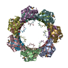


 Mass: 24.305 Da / Num. of mol.: 27 / Source method: obtained synthetically / Formula: Mg
 Mass: 18.015 Da / Num. of mol.: 91 / Source method: isolated from a natural source / Formula: H2O
 Sample preparation
 / Beamline: 31-ID / Wavelength: 0.979 Å
 Processing