 Movie
Movie Controller

[English] 日本語
 Yorodumi
- PDB-3blv: Yeast Isocitrate Dehydrogenase with Citrate Bound in the Regulato... -
+ Open data

- Basic information
| Entry | Database: PDB / ID: 3blv | ||||||
|---|---|---|---|---|---|---|---|
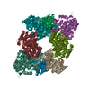
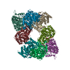
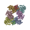
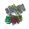
| Title | Yeast Isocitrate Dehydrogenase with Citrate Bound in the Regulatory Subunits | ||||||
 Components Components |
| ||||||
 Keywords Keywords | OXIDOREDUCTASE / TCA cycle / oxidative metabolism / allostery / dehydrogenase / decarboxylase / Allosteric enzyme / Magnesium / Manganese / Metal-binding / Mitochondrion / NAD / RNA-binding / Transit peptide / Tricarboxylic acid cycle / Phosphoprotein | ||||||
| Function / homology |  Function and homology information Function and homology informationCitric acid cycle (TCA cycle) / isocitrate dehydrogenase complex (NAD+) / isocitrate dehydrogenase (NAD+) / isocitrate dehydrogenase (NAD+) activity / isocitrate metabolic process / : / mitochondrial nucleoid / tricarboxylic acid cycle / mitochondrial intermembrane space / NAD binding ...Citric acid cycle (TCA cycle) / isocitrate dehydrogenase complex (NAD+) / isocitrate dehydrogenase (NAD+) / isocitrate dehydrogenase (NAD+) activity / isocitrate metabolic process / : / mitochondrial nucleoid / tricarboxylic acid cycle / mitochondrial intermembrane space / NAD binding / mitochondrial matrix / magnesium ion binding / mitochondrion / RNA binding / cytosol Similarity search - Function | ||||||
| Biological species |  | ||||||
| Method |  X-RAY DIFFRACTION / SYNCHROTRON / MAD, MOLECULAR REPLACEMENT / Resolution: 3.2 Å X-RAY DIFFRACTION / SYNCHROTRON / MAD, MOLECULAR REPLACEMENT / Resolution: 3.2 Å | ||||||
 Authors Authors | Taylor, A.B. / Hu, G. / Hart, P.J. / McAlister-Henn, L. | ||||||
 Citation Citation | Journal: J.Biol.Chem. / Year: 2008 Title: Allosteric Motions in Structures of Yeast NAD+-specific Isocitrate Dehydrogenase. Authors: Taylor, A.B. / Hu, G. / Hart, P.J. / McAlister-Henn, L. | ||||||
| History |
|
- Structure visualization
| Structure viewer | Molecule: MolmilJmol/JSmol |
|---|
- Downloads & links
-Download
| PDBx/mmCIF format | 3blv.cif.gz | 511.4 KB | Display | PDBx/mmCIF format |
|---|---|---|---|---|
| PDB format | pdb3blv.ent.gz | 422.7 KB | Display | PDB format |
| PDBx/mmJSON format | 3blv.json.gz | Tree view | PDBx/mmJSON format | |
| Others |  Other downloads Other downloads |
-Validation report
| Arichive directory | https://data.pdbj.org/pub/pdb/validation_reports/bl/3blvftp://data.pdbj.org/pub/pdb/validation_reports/bl/3blv | HTTPS FTP |
|---|
-Related structure data
| Related structure data | 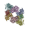 3blwC  3blxC 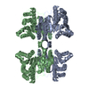 1wpwS C: citing same article ( S: Starting model for refinement |
|---|---|
| Similar structure data |


-Links
 PDBj
PDBj


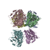
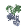
- Assembly
| Deposited unit | 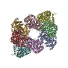
| ||||||||
|---|---|---|---|---|---|---|---|---|---|
| 1 |
| ||||||||
| 2 | 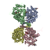
| ||||||||
| 3 | 
| ||||||||
| Unit cell |
|
-Components
| #1: Protein | Mass: 39095.336 Da / Num. of mol.: 4 Source method: isolated from a genetically manipulated source Source: (gene. exp.) Gene: IDH1 / Plasmid: pET15b / Production host:  References: UniProt: P28834, isocitrate dehydrogenase (NAD+) #2: Protein | Mass: 37979.711 Da / Num. of mol.: 4 Source method: isolated from a genetically manipulated source Source: (gene. exp.) Gene: IDH2 / Plasmid: pET15b / Production host: References: UniProt: P28241, isocitrate dehydrogenase (NAD+) #3: Chemical | ChemComp-FLC /   Mass: 189.100 Da / Num. of mol.: 4 / Source method: obtained synthetically / Formula: C6H5O7 Mass: 189.100 Da / Num. of mol.: 4 / Source method: obtained synthetically / Formula: C6H5O7Has protein modification | Y | |
|---|
-Experimental details
-Experiment
| Experiment | Method: X-RAY DIFFRACTION / Number of used crystals: 1 |
|---|
- Sample preparation
| Crystal | Density Matthews: 2.83 Å3/Da / Density % sol: 56.56 % Description: THE STRUCTURE FACTOR FILE CONTAINS FRIEDEL PAIRS |
|---|---|
| Crystal grow | Temperature: 295 K / Method: vapor diffusion, hanging drop / pH: 9.5 Details: 1.1 M sodium citrate, 0.1 M CHES, 15% ethylene glycol, pH 9.5, VAPOR DIFFUSION, HANGING DROP, temperature 295K |
-Data collection
| Diffraction | Mean temperature: 100 K | |||||||||
|---|---|---|---|---|---|---|---|---|---|---|
| Diffraction source | Source: SYNCHROTRON / Site: ALS  / Beamline: 5.0.2 / Wavelength: 0.9791, 0.9796 / Beamline: 5.0.2 / Wavelength: 0.9791, 0.9796 | |||||||||
| Detector | Type: ADSC QUANTUM 315 / Detector: CCD / Date: Nov 7, 2003 | |||||||||
| Radiation | Protocol: MAD / Monochromatic (M) / Laue (L): M / Scattering type: x-ray | |||||||||
| Radiation wavelength |
| |||||||||
| Reflection | Resolution: 3.2→50 Å / Num. obs: 109531 / % possible obs: 96.8 % / Redundancy: 3.1 % / Biso Wilson estimate: 68.6 Å2 / Rsym value: 0.101 / Net I/σ(I): 11 | |||||||||
| Reflection shell | Resolution: 3.2→3.31 Å / Redundancy: 3 % / Mean I/σ(I) obs: 2.1 / Num. unique all: 11104 / Rsym value: 0.556 / % possible all: 97.9 |
- Processing
| Software |
| ||||||||||||||||||||
|---|---|---|---|---|---|---|---|---|---|---|---|---|---|---|---|---|---|---|---|---|---|
| Refinement | Method to determine structure: MAD, MOLECULAR REPLACEMENT Starting model: PDB entry 1WPW Resolution: 3.2→49.346 Å / Isotropic thermal model: isotropic / Cross valid method: THROUGHOUT / Details: THE FRIEDEL PAIRS WERE USED FOR PHASING
| ||||||||||||||||||||
| Displacement parameters | Biso mean: 73.9 Å2
| ||||||||||||||||||||
| Refinement step | Cycle: LAST / Resolution: 3.2→49.346 Å
| ||||||||||||||||||||
| Refine LS restraints |
| ||||||||||||||||||||
| LS refinement shell | Resolution: 3.2→3.2364 Å
|
