 Movie
Movie Controller

[English] 日本語
 Yorodumi
- PDB-3bjv: The Crystal Structure of a putative PTS IIA(PtxA) from Streptococ... -
+ Open data

- Basic information
| Entry | Database: PDB / ID: 3bjv | ||||||
|---|---|---|---|---|---|---|---|
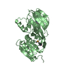
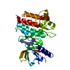
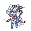
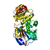
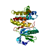
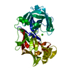
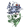
| Title | The Crystal Structure of a putative PTS IIA(PtxA) from Streptococcus mutans | ||||||
 Components Components | RmpA | ||||||
 Keywords Keywords | TRANSFERASE / alpha/beta three layer sandwich | ||||||
| Function / homology |  Function and homology information Function and homology informationphosphoenolpyruvate-dependent sugar phosphotransferase system / kinase activity / cytoplasm Similarity search - Function | ||||||
| Biological species |  Streptococcus mutans (bacteria) Streptococcus mutans (bacteria) | ||||||
| Method |  X-RAY DIFFRACTION / MOLECULAR REPLACEMENT / molecular replacement / Resolution: 2.4 Å X-RAY DIFFRACTION / MOLECULAR REPLACEMENT / molecular replacement / Resolution: 2.4 Å | ||||||
 Authors Authors | Lei, J. / Liang, Y.H. / Su, X.D. | ||||||
 Citation Citation | Journal: J.Mol.Biol. / Year: 2009 Title: Crystal structures of phosphotransferase system enzymes PtxB (IIB(Asc)) and PtxA (IIA(Asc)) from Streptococcus mutans Authors: Lei, J. / Li, L.-F. / Su, X.-D. | ||||||
| History |
|
- Structure visualization
| Structure viewer | Molecule: MolmilJmol/JSmol |
|---|
- Downloads & links
-Download
| PDBx/mmCIF format | 3bjv.cif.gz | 43.4 KB | Display | PDBx/mmCIF format |
|---|---|---|---|---|
| PDB format | pdb3bjv.ent.gz | 29.7 KB | Display | PDB format |
| PDBx/mmJSON format | 3bjv.json.gz | Tree view | PDBx/mmJSON format | |
| Others |  Other downloads Other downloads |
-Validation report
| Arichive directory | https://data.pdbj.org/pub/pdb/validation_reports/bj/3bjvftp://data.pdbj.org/pub/pdb/validation_reports/bj/3bjv | HTTPS FTP |
|---|
-Related structure data
| Related structure data |  3czcC  2oqtS S: Starting model for refinement C: citing same article ( |
|---|---|
| Similar structure data |


-Links
 PDBj
PDBj- Assembly
| Deposited unit | 
| ||||||||
|---|---|---|---|---|---|---|---|---|---|
| 1 | 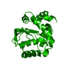
| ||||||||
| Unit cell |
|
-Components
| #1: Protein | Mass: 17696.219 Da / Num. of mol.: 1 Source method: isolated from a genetically manipulated source Source: (gene. exp.) Streptococcus mutans (bacteria) / Strain: UA159 / Gene: ptxA / Plasmid: pET28a / Species (production host): Escherichia coli / Production host: References: UniProt: Q93DA9, protein-Npi-phosphohistidine-sugar phosphotransferase |
|---|---|
| #2: Water | ChemComp-HOH /  Mass: 18.015 Da / Num. of mol.: 45 / Source method: isolated from a natural source / Formula: H2O Mass: 18.015 Da / Num. of mol.: 45 / Source method: isolated from a natural source / Formula: H2O |
-Experimental details
-Experiment
| Experiment | Method: X-RAY DIFFRACTION / Number of used crystals: 1 |
|---|
- Sample preparation
| Crystal | Density Matthews: 2.05 Å3/Da / Density % sol: 40.07 % |
|---|---|
| Crystal grow | Temperature: 289 K / Method: vapor diffusion / pH: 7.9 Details: 0.1M Tris-Hcl, 1.9-2.1M (NH4)2SO4, pH 7.9, Vapor diffusion, temperature 289K |
-Data collection
| Diffraction | Mean temperature: 293 K |
|---|---|
| Diffraction source | Source: ROTATING ANODE / Type: BRUKER AXS MICROSTAR-H / Wavelength: 1.5418 Å |
| Detector | Type: BRUKER SMART 6000 / Detector: CCD / Date: Dec 2, 2007 |
| Radiation | Monochromator: multilayer mirror / Protocol: SINGLE WAVELENGTH / Monochromatic (M) / Laue (L): M / Scattering type: x-ray |
| Radiation wavelength | Wavelength: 1.5418 Å / Relative weight: 1 |
| Reflection | Resolution: 2.4→40.65 Å / Num. all: 6206 / Num. obs: 6026 / % possible obs: 97.1 % / Redundancy: 9.3 % / Biso Wilson estimate: 32 Å2 / Rmerge(I) obs: 0.0717 / Rsym value: 0.0717 / Net I/σ(I): 10.08 |
| Reflection shell | Resolution: 2.4→2.54 Å / Redundancy: 9 % / Rmerge(I) obs: 0.1297 / Mean I/σ(I) obs: 0.1297 / Num. unique all: 897 / Rsym value: 0.1297 / % possible all: 85.1 |
-Phasing
| Phasing | Method: molecular replacement | |||||||||
|---|---|---|---|---|---|---|---|---|---|---|
| Phasing MR |
|
- Processing
| Software |
| ||||||||||||||||||||||||||||||||||||||||||||||||||||||||||||||||||||||||||||||||||||||||||
|---|---|---|---|---|---|---|---|---|---|---|---|---|---|---|---|---|---|---|---|---|---|---|---|---|---|---|---|---|---|---|---|---|---|---|---|---|---|---|---|---|---|---|---|---|---|---|---|---|---|---|---|---|---|---|---|---|---|---|---|---|---|---|---|---|---|---|---|---|---|---|---|---|---|---|---|---|---|---|---|---|---|---|---|---|---|---|---|---|---|---|---|
| Refinement | Method to determine structure: MOLECULAR REPLACEMENT Starting model: PDB ENTRY 2OQT Resolution: 2.4→27.28 Å / Cor.coef. Fo:Fc: 0.954 / Cor.coef. Fo:Fc free: 0.883 / SU B: 7.265 / SU ML: 0.173 / Cross valid method: THROUGHOUT / σ(F): 0 / ESU R: 1.114 / ESU R Free: 0.302 / Stereochemistry target values: MAXIMUM LIKELIHOOD / Details: HYDROGENS HAVE BEEN ADDED IN THE RIDING POSITIONS
| ||||||||||||||||||||||||||||||||||||||||||||||||||||||||||||||||||||||||||||||||||||||||||
| Solvent computation | Ion probe radii: 0.8 Å / Shrinkage radii: 0.8 Å / VDW probe radii: 1.4 Å / Solvent model: MASK | ||||||||||||||||||||||||||||||||||||||||||||||||||||||||||||||||||||||||||||||||||||||||||
| Displacement parameters | Biso mean: 15.719 Å2
| ||||||||||||||||||||||||||||||||||||||||||||||||||||||||||||||||||||||||||||||||||||||||||
| Refine analyze | Luzzati coordinate error obs: 0.232 Å | ||||||||||||||||||||||||||||||||||||||||||||||||||||||||||||||||||||||||||||||||||||||||||
| Refinement step | Cycle: LAST / Resolution: 2.4→27.28 Å
| ||||||||||||||||||||||||||||||||||||||||||||||||||||||||||||||||||||||||||||||||||||||||||
| Refine LS restraints |
| ||||||||||||||||||||||||||||||||||||||||||||||||||||||||||||||||||||||||||||||||||||||||||
| LS refinement shell | Resolution: 2.397→2.459 Å / Total num. of bins used: 20
|
