 Movie
Movie Controller

[English] 日本語
 Yorodumi
- PDB-2ans: ADIPOCYTE LIPID BINDING PROTEIN COMPLEXED WITH 1-ANILINO-8-NAPHTH... -
+ Open data

- Basic information
| Entry | Database: PDB / ID: 2ans | ||||||
|---|---|---|---|---|---|---|---|
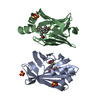
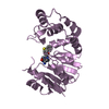
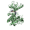
| Title | ADIPOCYTE LIPID BINDING PROTEIN COMPLEXED WITH 1-ANILINO-8-NAPHTHALENE SULFONATE | ||||||
 Components Components | ADIPOCYTE LIPID-BINDING PROTEIN | ||||||
 Keywords Keywords | LIPID BINDING PROTEIN / LIPID-BINDING PROTEIN / FLUORESCENT PROBE | ||||||
| Function / homology |  Function and homology information Function and homology informationlipase activator activity / Triglyceride catabolism / lipid droplet formation / triglyceride catabolic process / hormone receptor binding / long-chain fatty acid transmembrane transporter activity / long-chain fatty acid binding / cellular response to lithium ion / white fat cell differentiation / long-chain fatty acid transport ...lipase activator activity / Triglyceride catabolism / lipid droplet formation / triglyceride catabolic process / hormone receptor binding / long-chain fatty acid transmembrane transporter activity / long-chain fatty acid binding / cellular response to lithium ion / white fat cell differentiation / long-chain fatty acid transport / fatty acid transport / lipid droplet / brown fat cell differentiation / fatty acid binding / cholesterol homeostasis / response to bacterium / fatty acid metabolic process / cellular response to tumor necrosis factor / positive regulation of inflammatory response / positive regulation of cold-induced thermogenesis / negative regulation of DNA-templated transcription / positive regulation of cell population proliferation / nucleoplasm / nucleus / cytoplasm / cytosol Similarity search - Function | ||||||
| Biological species |  | ||||||
| Method |  X-RAY DIFFRACTION / MOLECULAR REPLACEMENT / Resolution: 2.5 Å X-RAY DIFFRACTION / MOLECULAR REPLACEMENT / Resolution: 2.5 Å | ||||||
 Authors Authors | Ory, J.J. / Banaszak, L.J. | ||||||
 Citation Citation | Journal: Biophys.J. / Year: 1999 Title: Studies of the ligand binding reaction of adipocyte lipid binding protein using the fluorescent probe 1, 8-anilinonaphthalene-8-sulfonate. Authors: Ory, J.J. / Banaszak, L.J. | ||||||
| History |
|
- Structure visualization
| Structure viewer | Molecule: MolmilJmol/JSmol |
|---|
- Downloads & links
-Download
| PDBx/mmCIF format | 2ans.cif.gz | 63.5 KB | Display | PDBx/mmCIF format |
|---|---|---|---|---|
| PDB format | pdb2ans.ent.gz | 47.4 KB | Display | PDB format |
| PDBx/mmJSON format | 2ans.json.gz | Tree view | PDBx/mmJSON format | |
| Others |  Other downloads Other downloads |
-Validation report
| Arichive directory | https://data.pdbj.org/pub/pdb/validation_reports/an/2ansftp://data.pdbj.org/pub/pdb/validation_reports/an/2ans | HTTPS FTP |
|---|
-Related structure data
| Related structure data |  1libS S: Starting model for refinement |
|---|---|
| Similar structure data |


-Links
 PDBj
PDBj

- Assembly
| Deposited unit | 
| ||||||||
|---|---|---|---|---|---|---|---|---|---|
| 1 |
| ||||||||
| Unit cell |
| ||||||||
| Noncrystallographic symmetry (NCS) | NCS oper: (Code: given Matrix: (-0.99105, 0.13205, 0.019552), Vector: |
-Components
| #1: Protein | Mass: 14539.688 Da / Num. of mol.: 2 / Source method: isolated from a natural source / Source: (natural) #2: Chemical | ChemComp-CL / |   Mass: 35.453 Da / Num. of mol.: 1 / Source method: obtained synthetically / Formula: Cl Mass: 35.453 Da / Num. of mol.: 1 / Source method: obtained synthetically / Formula: Cl#3: Chemical |   Mass: 299.344 Da / Num. of mol.: 2 / Source method: obtained synthetically / Formula: C16H13NO3S Mass: 299.344 Da / Num. of mol.: 2 / Source method: obtained synthetically / Formula: C16H13NO3S#4: Water | ChemComp-HOH / |  Mass: 18.015 Da / Num. of mol.: 78 / Source method: isolated from a natural source / Formula: H2O Mass: 18.015 Da / Num. of mol.: 78 / Source method: isolated from a natural source / Formula: H2O |
|---|
-Experimental details
-Experiment
| Experiment | Method: X-RAY DIFFRACTION / Number of used crystals: 1 |
|---|
- Sample preparation
| Crystal | Density Matthews: 3.34 Å3/Da / Density % sol: 63 % | ||||||||||||||||||||
|---|---|---|---|---|---|---|---|---|---|---|---|---|---|---|---|---|---|---|---|---|---|
| Crystal grow | pH: 7.5 / Details: pH 7.5 | ||||||||||||||||||||
| Crystal grow | *PLUS Method: vapor diffusion, hanging drop | ||||||||||||||||||||
| Components of the solutions | *PLUS
|
-Data collection
| Diffraction | Mean temperature: 298 K |
|---|---|
| Diffraction source | Source: ROTATING ANODE / Type: RIGAKU RUH2R / Wavelength: 1.5418 |
| Detector | Type: SIEMENS / Detector: AREA DETECTOR |
| Radiation | Monochromator: GRAPHITE(002) / Monochromatic (M) / Laue (L): M / Scattering type: x-ray |
| Radiation wavelength | Wavelength: 1.5418 Å / Relative weight: 1 |
| Reflection | Resolution: 2.5→24 Å / Num. obs: 12287 / % possible obs: 95 % / Observed criterion σ(I): 0 / Redundancy: 6 % / Rsym value: 0.102 |
| Reflection shell | Resolution: 2.5→2.7 Å / Redundancy: 2.5 % / Rsym value: 0.312 / % possible all: 58 |
| Reflection | *PLUS Num. measured all: 74950 / Rmerge(I) obs: 0.102 |
| Reflection shell | *PLUS % possible obs: 58 % / Num. unique obs: 1263 / Rmerge(I) obs: 0.312 |
- Processing
| Software |
| ||||||||||||||||||||||||||||||||||||||||||||||||||||||||||||
|---|---|---|---|---|---|---|---|---|---|---|---|---|---|---|---|---|---|---|---|---|---|---|---|---|---|---|---|---|---|---|---|---|---|---|---|---|---|---|---|---|---|---|---|---|---|---|---|---|---|---|---|---|---|---|---|---|---|---|---|---|---|
| Refinement | Method to determine structure: MOLECULAR REPLACEMENT Starting model: 1LIB Resolution: 2.5→24 Å / Cross valid method: THROUGHOUT / σ(F): 0
| ||||||||||||||||||||||||||||||||||||||||||||||||||||||||||||
| Refinement step | Cycle: LAST / Resolution: 2.5→24 Å
| ||||||||||||||||||||||||||||||||||||||||||||||||||||||||||||
| Refine LS restraints |
| ||||||||||||||||||||||||||||||||||||||||||||||||||||||||||||
| Xplor file |
| ||||||||||||||||||||||||||||||||||||||||||||||||||||||||||||
| Software | *PLUS Name: X-PLOR / Version: 3.8 / Classification: refinement | ||||||||||||||||||||||||||||||||||||||||||||||||||||||||||||
| Refine LS restraints | *PLUS Type: x_angle_deg / Dev ideal: 1.581 |
