Resolution: 1.76→29.223 Å / Num. obs: 27863 / % possible obs: 99.4 % / Observed criterion σ(I): -3 / Redundancy: 10.35 % / Biso Wilson estimate: 22.32 Å2 / Rmerge(I) obs: 0.083 / Net I/σ(I): 13.14
Reflection shell
Resolution (Å)
Rmerge(I) obs
Mean I/σ(I) obs
Num. measured obs
Diffraction-ID
% possible all
1.76-1.82
0.806
2
26238
1
95.4
1.82-1.9
0.6
2.8
31636
1
100
1.9-1.98
0.436
4
26930
1
99.9
1.98-2.09
0.29
6.1
30581
1
100
2.09-2.22
0.193
8.8
28768
1
100
2.22-2.39
0.139
11.8
28798
1
100
2.39-2.63
0.101
15.3
29162
1
100
2.63-3.01
0.072
20
28919
1
99.9
3.01-3.78
0.05
27.6
28654
1
100
3.78-29.223
0.044
32.6
28499
1
99
-
Phasing
Phasing
Method: MAD
-
Processing
Software
Name
Version
Classification
NB
REFMAC
5.2.0019
refinement
PHENIX
refinement
SOLVE
phasing
MolProbity
3beta29
modelbuilding
XSCALE
datascaling
PDB_EXTRACT
3
dataextraction
ADSC
Quantum
datacollection
XDS
datareduction
Refinement
Method to determine structure: MAD / Resolution: 1.76→29.223 Å / Cor.coef. Fo:Fc: 0.967 / Cor.coef. Fo:Fc free: 0.956 / SU B: 3.797 / SU ML: 0.061 / TLS residual ADP flag: LIKELY RESIDUAL / Cross valid method: THROUGHOUT / σ(F): 0 / ESU R: 0.092 / ESU R Free: 0.091 Stereochemistry target values: MAXIMUM LIKELIHOOD WITH PHASES Details: 1. HYDROGENS HAVE BEEN ADDED IN THE RIDING POSITIONS. 2. A MET-INHIBITION PROTOCOL WAS USED FOR SELENOMETHIONINE INCORPORATION DURING PROTEIN EXPRESSION. THE OCCUPANCY OF THE SE ATOMS IN THE ...Details: 1. HYDROGENS HAVE BEEN ADDED IN THE RIDING POSITIONS. 2. A MET-INHIBITION PROTOCOL WAS USED FOR SELENOMETHIONINE INCORPORATION DURING PROTEIN EXPRESSION. THE OCCUPANCY OF THE SE ATOMS IN THE MSE RESIDUES WAS REDUCED TO 0.75 TO ACCOUNT FOR THE REDUCED SCATTERING POWER DUE TO PARTIAL S-MET INCORPORATION. 3. ATOM RECORD CONTAINS RESIDUAL B FACTORS ONLY. 4. 1,2-ETHANEDIOL MOLECULES FROM THE CRYSTALLIZATION CONDITIONS HAVE BEEN MODELED IN THE SOLVENT STRUCTURE.
Rfactor
Num. reflection
% reflection
Selection details
Rfree
0.19
1398
5 %
RANDOM
Rwork
0.162
-
-
-
obs
0.163
27782
99.88 %
-
Solvent computation
Ion probe radii: 0.8 Å / Shrinkage radii: 0.8 Å / VDW probe radii: 1.2 Å / Solvent model: MASK
Displacement parameters
Biso mean: 18.478 Å2
Baniso -1
Baniso -2
Baniso -3
1-
0.59 Å2
0.3 Å2
0 Å2
2-
-
0.59 Å2
0 Å2
3-
-
-
-0.89 Å2
Refinement step
Cycle: LAST / Resolution: 1.76→29.223 Å
Protein
Nucleic acid
Ligand
Solvent
Total
Num. atoms
1604
0
12
204
1820
Refine LS restraints
Refine-ID
Type
Dev ideal
Dev ideal target
Number
X-RAY DIFFRACTION
r_bond_refined_d
0.015
0.022
1728
X-RAY DIFFRACTION
r_bond_other_d
0.001
0.02
1133
X-RAY DIFFRACTION
r_angle_refined_deg
1.397
1.972
2360
X-RAY DIFFRACTION
r_angle_other_deg
0.984
3
2815
X-RAY DIFFRACTION
r_dihedral_angle_1_deg
4.968
5
242
X-RAY DIFFRACTION
r_dihedral_angle_2_deg
37.769
25.714
70
X-RAY DIFFRACTION
r_dihedral_angle_3_deg
12.681
15
301
X-RAY DIFFRACTION
r_dihedral_angle_4_deg
20.813
15
5
X-RAY DIFFRACTION
r_chiral_restr
0.092
0.2
279
X-RAY DIFFRACTION
r_gen_planes_refined
0.005
0.02
1960
X-RAY DIFFRACTION
r_gen_planes_other
0.001
0.02
323
X-RAY DIFFRACTION
r_nbd_refined
0.213
0.2
351
X-RAY DIFFRACTION
r_nbd_other
0.192
0.2
1195
X-RAY DIFFRACTION
r_nbtor_refined
0.174
0.2
847
X-RAY DIFFRACTION
r_nbtor_other
0.089
0.2
857
X-RAY DIFFRACTION
r_xyhbond_nbd_refined
0.177
0.2
165
X-RAY DIFFRACTION
r_symmetry_vdw_refined
0.169
0.2
14
X-RAY DIFFRACTION
r_symmetry_vdw_other
0.158
0.2
24
X-RAY DIFFRACTION
r_symmetry_hbond_refined
0.219
0.2
18
X-RAY DIFFRACTION
r_mcbond_it
1.862
3
1185
X-RAY DIFFRACTION
r_mcbond_other
0.461
3
456
X-RAY DIFFRACTION
r_mcangle_it
2.502
5
1790
X-RAY DIFFRACTION
r_scbond_it
4.785
8
671
X-RAY DIFFRACTION
r_scangle_it
6.046
11
556
LS refinement shell
Resolution: 1.76→1.81 Å / Total num. of bins used: 20
Rfactor
Num. reflection
% reflection
Rfree
0.273
109
-
Rwork
0.218
1869
-
all
-
1978
-
obs
-
-
98.95 %
Refinement TLS params.
Method: refined / Refine-ID: X-RAY DIFFRACTION
ID
L11 (°2)
L12 (°2)
L13 (°2)
L22 (°2)
L23 (°2)
L33 (°2)
S11 (Å °)
S12 (Å °)
S13 (Å °)
S21 (Å °)
S22 (Å °)
S23 (Å °)
S31 (Å °)
S32 (Å °)
S33 (Å °)
T11 (Å2)
T12 (Å2)
T13 (Å2)
T22 (Å2)
T23 (Å2)
T33 (Å2)
Origin x (Å)
Origin y (Å)
Origin z (Å)
1
1.1771
-0.0202
0.3503
1.5381
-0.711
1.0973
0.0337
0.0231
-0.0758
-0.064
0.1154
0.1004
0.0917
-0.1528
-0.1491
-0.0127
-0.0107
-0.0453
-0.0462
0.0377
-0.0284
-8.4506
65.3131
23.1685
2
1.2614
0.421
-0.0549
1.2876
-0.3008
1.4684
0.0464
-0.1459
-0.0033
0.249
0.0081
-0.0895
-0.098
0.003
-0.0546
0.0152
-0.0024
-0.0538
-0.0514
0.014
-0.0479
2.6618
72.818
32.5279
3
0.9434
0.0503
0.4371
1.0057
-0.1668
0.8699
0.0701
0.1454
-0.0353
0.0364
-0.1076
-0.2168
0.092
0.1515
0.0375
-0.0278
0.0201
-0.0261
-0.0158
0.0127
0.005
11.1304
73.6973
18.9981
Refinement TLS group
ID
Refine-ID
Refine TLS-ID
Auth asym-ID
Label asym-ID
Auth seq-ID
Label seq-ID
1
X-RAY DIFFRACTION
1
A
A
1 - 88
20 - 107
2
X-RAY DIFFRACTION
2
A
A
89 - 152
108 - 171
3
X-RAY DIFFRACTION
3
A
A
153 - 214
172 - 233
+
About Yorodumi
-
News
-
Feb 9, 2022. New format data for meta-information of EMDB entries
New format data for meta-information of EMDB entries
Version 3 of the EMDB header file is now the official format.
The previous official version 1.9 will be removed from the archive.
In the structure databanks used in Yorodumi, some data are registered as the other names, "COVID-19 virus" and "2019-nCoV". Here are the details of the virus and the list of structure data.
Jan 31, 2019. EMDB accession codes are about to change! (news from PDBe EMDB page)
EMDB accession codes are about to change! (news from PDBe EMDB page)
The allocation of 4 digits for EMDB accession codes will soon come to an end. Whilst these codes will remain in use, new EMDB accession codes will include an additional digit and will expand incrementally as the available range of codes is exhausted. The current 4-digit format prefixed with “EMD-” (i.e. EMD-XXXX) will advance to a 5-digit format (i.e. EMD-XXXXX), and so on. It is currently estimated that the 4-digit codes will be depleted around Spring 2019, at which point the 5-digit format will come into force.
The EM Navigator/Yorodumi systems omit the EMD- prefix.
Related info.:Q: What is EMD? / ID/Accession-code notation in Yorodumi/EM Navigator
Yorodumi is a browser for structure data from EMDB, PDB, SASBDB, etc.
This page is also the successor to EM Navigator detail page, and also detail information page/front-end page for Omokage search.
The word "yorodu" (or yorozu) is an old Japanese word meaning "ten thousand". "mi" (miru) is to see.
Related info.:EMDB / PDB / SASBDB / Comparison of 3 databanks / Yorodumi Search / Aug 31, 2016. New EM Navigator & Yorodumi / Yorodumi Papers / Jmol/JSmol / Function and homology information / Changes in new EM Navigator and Yorodumi
 Movie
Movie Controller

 Yorodumi
 Open data
 Basic information
 Components
 Keywords
 Function and homology information
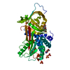
 Shewanella loihica PV-4 (bacteria)
 X-RAY DIFFRACTION /
  Authors
 Citation
 Structure visualization
 Downloads & links
 Other downloads
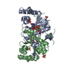


 PDBj
PDBj

 Assembly
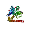

 Mass: 62.068 Da / Num. of mol.: 3 / Source method: obtained synthetically / Formula: C2H6O2
 Mass: 18.015 Da / Num. of mol.: 204 / Source method: isolated from a natural source / Formula: H2O
 Sample preparation
 / Beamline: 8.2.2 / Wavelength: 0.9537, 0.9798, 0.9796
 Processing