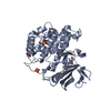
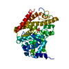
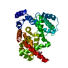
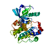
Entry Database : PDB / ID : 3beaTitle cFMS tyrosine kinase (tie2 KID) in complex with a pyrimidinopyridone inhibitor Macrophage colony-stimulating factor 1 receptor Keywords / / / / / / / / / / / / / Function / homology Function Domain/homology Component
/ / / / / / / / / / / / / / / / / / / / / / / / / / / / / / / / / / / / / / / / / / / / / / / / / / / / / / / / / / / / / / / / / / / / / / / / / / / / / / / / / / / / / / / / / / / / / / / / / / / / / / / / / / / / / / / / / / / / / / / / / / / / / / / / / / / / / / / / / / / / / / / / / / / / / / / / / / / / / / / / / / / / Biological species Homo sapiens (human)Method / / / Resolution : 2.02 Å Authors Schubert, C. Journal : Bioorg.Med.Chem.Lett. / Year : 2008Title : Design and synthesis of a pyrido[2,3-d]pyrimidin-5-one class of anti-inflammatory FMS inhibitors.Authors : Huang, H. / Hutta, D.A. / Hu, H. / DesJarlais, R.L. / Schubert, C. / Petrounia, I.P. / Chaikin, M.A. / Manthey, C.L. / Player, M.R. History Deposition Nov 16, 2007 Deposition site / Processing site Revision 1.0 Jul 15, 2008 Provider / Type Revision 1.1 Jul 13, 2011 Group Revision 1.2 Aug 2, 2017 Group / Category Revision 1.3 Oct 20, 2021 Group / Derived calculations / Category / struct_ref_seq_dif / struct_siteItem _database_2.pdbx_DOI / _database_2.pdbx_database_accession ... _database_2.pdbx_DOI / _database_2.pdbx_database_accession / _struct_ref_seq_dif.details / _struct_site.pdbx_auth_asym_id / _struct_site.pdbx_auth_comp_id / _struct_site.pdbx_auth_seq_id Revision 1.4 Aug 30, 2023 Group / Refinement descriptionCategory / chem_comp_bond / pdbx_initial_refinement_model
Show all Show less
 Movie
Movie Controller

 Yorodumi
 Open data
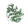
 Basic information
 Components
 Keywords
 Function and homology information
 Homo sapiens (human)
 X-RAY DIFFRACTION /
  Authors
 Citation
 Structure visualization
 Downloads & links
 Other downloads


 PDBj
PDBj


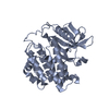
 Assembly

 Spodoptera frugiperda (fall armyworm)
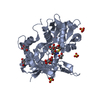
 Mass: 96.063 Da / Num. of mol.: 3 / Source method: obtained synthetically / Formula: SO4
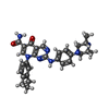
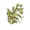
 Mass: 509.602 Da / Num. of mol.: 1 / Source method: obtained synthetically / Formula: C29H31N7O2
 Mass: 18.015 Da / Num. of mol.: 169 / Source method: isolated from a natural source / Formula: H2O
 Sample preparation
 / Beamline: 17-BM / Wavelength: 1 Å
 Processing