 Movie
Movie Controller


+ Open data

- Basic information
| Entry | Database: PDB / ID: 3bck | ||||||
|---|---|---|---|---|---|---|---|
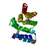
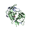
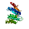
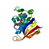
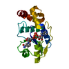
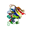
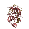
| Title | Crystal Structure of Staphylococcus aureus DsbA T153V | ||||||
 Components Components | Disulfide bond protein A | ||||||
 Keywords Keywords | OXIDOREDUCTASE / thiol-disulfide oxidoreductase / redox protein / protein folding / redox active centre | ||||||
| Function / homology |  Function and homology information Function and homology information | ||||||
| Biological species |   Staphylococcus aureus (bacteria) Staphylococcus aureus (bacteria) | ||||||
| Method |  X-RAY DIFFRACTION / MOLECULAR REPLACEMENT / Resolution: 1.95 Å X-RAY DIFFRACTION / MOLECULAR REPLACEMENT / Resolution: 1.95 Å | ||||||
 Authors Authors | Heras, B. / Thony-Meyer, L. / Martin, J.L. | ||||||
 Citation Citation | Journal: J.Biol.Chem. / Year: 2008 Title: Staphylococcus aureus DsbA Does Not Have a Destabilizing Disulfide: A NEW PARADIGM FOR BACTERIAL OXIDATIVE FOLDING Authors: Heras, B. / Kurz, M. / Jarrott, R. / Shouldice, S.R. / Frei, P. / Robin, G. / Cemazar, M. / Thony-Meyer, L. / Glockshuber, R. / Martin, J.L. | ||||||
| History |
|
- Structure visualization
| Structure viewer | Molecule: MolmilJmol/JSmol |
|---|
- Downloads & links
-Download
| PDBx/mmCIF format | 3bck.cif.gz | 51 KB | Display | PDBx/mmCIF format |
|---|---|---|---|---|
| PDB format | pdb3bck.ent.gz | 35.6 KB | Display | PDB format |
| PDBx/mmJSON format | 3bck.json.gz | Tree view | PDBx/mmJSON format | |
| Others |  Other downloads Other downloads |
-Validation report
| Summary document | 3bck_validation.pdf.gz | 420.1 KB | Display | wwPDB validaton report |
|---|---|---|---|---|
| Full document | 3bck_full_validation.pdf.gz | 421.2 KB | Display | |
| Data in XML | 3bck_validation.xml.gz | 9.8 KB | Display | |
| Data in CIF | 3bck_validation.cif.gz | 13.6 KB | Display | |
| Arichive directory | https://data.pdbj.org/pub/pdb/validation_reports/bc/3bckftp://data.pdbj.org/pub/pdb/validation_reports/bc/3bck | HTTPS FTP |
-Related structure data
| Related structure data |  3bciSC 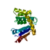 3bd2C S: Starting model for refinement C: citing same article ( |
|---|---|
| Similar structure data |


-Links
 PDBj
PDBj


- Assembly
| Deposited unit | 
| ||||||||
|---|---|---|---|---|---|---|---|---|---|
| 1 |
| ||||||||
| Unit cell |
|
-Components
| #1: Protein | Mass: 21808.006 Da / Num. of mol.: 1 / Fragment: residues in database 24-199 / Mutation: T153V Source method: isolated from a genetically manipulated source Source: (gene. exp.) Staphylococcus aureus (bacteria) / Strain: BB270 / Gene: AAG41993 / Plasmid: pET21a / Production host: |
|---|---|
| #2: Water | ChemComp-HOH /  Mass: 18.015 Da / Num. of mol.: 154 / Source method: isolated from a natural source / Formula: H2O Mass: 18.015 Da / Num. of mol.: 154 / Source method: isolated from a natural source / Formula: H2O |
| Has protein modification | Y |
-Experimental details
-Experiment
| Experiment | Method: X-RAY DIFFRACTION / Number of used crystals: 1 |
|---|
- Sample preparation
| Crystal | Density Matthews: 3.18378 Å3/Da / Density % sol: 65.86 % |
|---|---|
| Crystal grow | Temperature: 293 K / Method: vapor diffusion / Details: 28-30% PEG 3350, VAPOR DIFFUSION, temperature 293K |
-Data collection
| Diffraction | Mean temperature: 100 K |
|---|---|
| Diffraction source | Source: ROTATING ANODE / Type: RIGAKU FR-E+ SUPERBRIGHT / Wavelength: 1.54 Å |
| Detector | Type: RIGAKU RAXIS IV / Detector: IMAGE PLATE / Date: Oct 3, 2006 / Details: Osmic Confocal Max-Flux optics |
| Radiation | Monochromator: Osmic Confocal MaxFlux / Protocol: SINGLE WAVELENGTH / Monochromatic (M) / Laue (L): M / Scattering type: x-ray |
| Radiation wavelength | Wavelength: 1.54 Å / Relative weight: 1 |
| Reflection | Resolution: 1.95→62.43 Å / Num. all: 19271 / Num. obs: 19271 / % possible obs: 96.8 % / Observed criterion σ(F): 0 / Redundancy: 5.06 % / Rmerge(I) obs: 0.066 / Net I/σ(I): 9.3 |
| Reflection shell | Resolution: 1.95→2.02 Å / Redundancy: 4.07 % / Rmerge(I) obs: 0.523 / Mean I/σ(I) obs: 2.1 / Num. unique all: 1817 / % possible all: 92 |
- Processing
| Software |
| |||||||||||||||||||||||||
|---|---|---|---|---|---|---|---|---|---|---|---|---|---|---|---|---|---|---|---|---|---|---|---|---|---|---|
| Refinement | Method to determine structure: MOLECULAR REPLACEMENT Starting model: 3BCI Resolution: 1.95→62.43 Å / Isotropic thermal model: anisotropic / Cross valid method: THROUGHOUT / σ(F): 0 / Stereochemistry target values: Engh & Huber
| |||||||||||||||||||||||||
| Displacement parameters | Biso mean: 44.9 Å2
| |||||||||||||||||||||||||
| Refine analyze |
| |||||||||||||||||||||||||
| Refinement step | Cycle: LAST / Resolution: 1.95→62.43 Å
| |||||||||||||||||||||||||
| Refine LS restraints |
| |||||||||||||||||||||||||
| LS refinement shell | Resolution: 1.95→2.07 Å / Rfactor Rfree error: 0.035
| |||||||||||||||||||||||||
| Xplor file |
|
