 Movie
Movie Controller


+ Open data

- Basic information
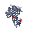
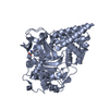
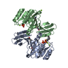
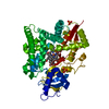
| Entry | Database: PDB / ID: 1bed | ||||||
|---|---|---|---|---|---|---|---|
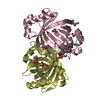
| Title | STRUCTURE OF DISULFIDE OXIDOREDUCTASE | ||||||
 Components Components | DSBA OXIDOREDUCTASE | ||||||
 Keywords Keywords | OXIDOREDUCTASE / TCPG / PROTEIN DISULFIDE ISOMERASE / DISULFIDE OXIDOREDUCTASE | ||||||
| Function / homology |  Function and homology information Function and homology informationprotein disulfide isomerase activity / cellular response to antibiotic / protein-disulfide reductase activity / outer membrane-bounded periplasmic space Similarity search - Function | ||||||
| Biological species |   Vibrio cholerae (bacteria) Vibrio cholerae (bacteria) | ||||||
| Method |  X-RAY DIFFRACTION / MOLECULAR REPLACEMENT / Resolution: 2 Å X-RAY DIFFRACTION / MOLECULAR REPLACEMENT / Resolution: 2 Å | ||||||
 Authors Authors | Hu, S.-H. / Martin, J.L. | ||||||
 Citation Citation | Journal: J.Mol.Biol. / Year: 1997 Title: Structure of TcpG, the DsbA protein folding catalyst from Vibrio cholerae. Authors: Hu, S.H. / Peek, J.A. / Rattigan, E. / Taylor, R.K. / Martin, J.L. #1: Journal: Proc.Natl.Acad.Sci.USA / Year: 1992Title: Characterization of a Periplasmic Thiol:Disulfide Interchange Protein Required for the Functional Maturation of Secreted Virulence Factors of Vibrio Cholerae Authors: Peek, J.A. / Taylor, R.K. | ||||||
| History |
|
- Structure visualization
| Structure viewer | Molecule: MolmilJmol/JSmol |
|---|
- Downloads & links
-Download
| PDBx/mmCIF format | 1bed.cif.gz | 47.8 KB | Display | PDBx/mmCIF format |
|---|---|---|---|---|
| PDB format | pdb1bed.ent.gz | 33.8 KB | Display | PDB format |
| PDBx/mmJSON format | 1bed.json.gz | Tree view | PDBx/mmJSON format | |
| Others |  Other downloads Other downloads |
-Validation report
| Arichive directory | https://data.pdbj.org/pub/pdb/validation_reports/be/1bedftp://data.pdbj.org/pub/pdb/validation_reports/be/1bed | HTTPS FTP |
|---|
-Related structure data
| Related structure data |  1dsbS S: Starting model for refinement |
|---|---|
| Similar structure data |


-Links
 PDBj
PDBj- Assembly
| Deposited unit | 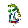
| ||||||||
|---|---|---|---|---|---|---|---|---|---|
| 1 | 
| ||||||||
| Unit cell |
|
-Components
| #1: Protein | Mass: 20523.418 Da / Num. of mol.: 1 / Source method: isolated from a natural source / Source: (natural) Vibrio cholerae (bacteria) / References: UniProt: P32557 |
|---|---|
| #2: Water | ChemComp-HOH /  Mass: 18.015 Da / Num. of mol.: 53 / Source method: isolated from a natural source / Formula: H2O Mass: 18.015 Da / Num. of mol.: 53 / Source method: isolated from a natural source / Formula: H2O |
| Has protein modification | Y |
-Experimental details
-Experiment
| Experiment | Method: X-RAY DIFFRACTION / Number of used crystals: 1 |
|---|
- Sample preparation
| Crystal | Density Matthews: 2.37 Å3/Da / Density % sol: 48 % | ||||||||||||||||||||||||||||||||||||||||||
|---|---|---|---|---|---|---|---|---|---|---|---|---|---|---|---|---|---|---|---|---|---|---|---|---|---|---|---|---|---|---|---|---|---|---|---|---|---|---|---|---|---|---|---|
| Crystal grow | pH: 6.5 / Details: 10-13% EG 4K, 0.1M MES, PH 6.0-6.5 / PH range: 6.0-6.5 | ||||||||||||||||||||||||||||||||||||||||||
| Crystal grow | *PLUS Temperature: 20 ℃ / pH: 6.8 / Method: vapor diffusion, hanging drop | ||||||||||||||||||||||||||||||||||||||||||
| Components of the solutions | *PLUS
|
-Data collection
| Diffraction | Mean temperature: 290 K |
|---|---|
| Diffraction source | Source: ROTATING ANODE / Type: RIGAKU RUH2R / Wavelength: 1.5418 |
| Detector | Type: RIGAKU RAXIS IIC / Detector: IMAGE PLATE / Date: Aug 17, 1994 |
| Radiation | Monochromator: GRAPHITE(002) / Monochromatic (M) / Laue (L): M / Scattering type: x-ray |
| Radiation wavelength | Wavelength: 1.5418 Å / Relative weight: 1 |
| Reflection | Resolution: 2→40 Å / Num. obs: 12974 / % possible obs: 98.2 % / Observed criterion σ(I): 0 / Redundancy: 5.7 % / Biso Wilson estimate: 31 Å2 / Rmerge(I) obs: 0.062 / Net I/σ(I): 13.1 |
| Reflection shell | Resolution: 2→2.07 Å / Redundancy: 2.9 % / Rmerge(I) obs: 0.341 / Mean I/σ(I) obs: 3.4 / % possible all: 91.7 |
| Reflection | *PLUS Num. measured all: 60945 |
| Reflection shell | *PLUS % possible obs: 91.7 % |
- Processing
| Software |
| ||||||||||||||||||||||||||||||||||||||||||||||||||||||||||||||||||||||||||||||||
|---|---|---|---|---|---|---|---|---|---|---|---|---|---|---|---|---|---|---|---|---|---|---|---|---|---|---|---|---|---|---|---|---|---|---|---|---|---|---|---|---|---|---|---|---|---|---|---|---|---|---|---|---|---|---|---|---|---|---|---|---|---|---|---|---|---|---|---|---|---|---|---|---|---|---|---|---|---|---|---|---|---|
| Refinement | Method to determine structure: MOLECULAR REPLACEMENT Starting model: PDB ENTRY 1DSB Resolution: 2→20 Å / Cross valid method: THROUGHOUT / σ(F): 2
| ||||||||||||||||||||||||||||||||||||||||||||||||||||||||||||||||||||||||||||||||
| Displacement parameters | Biso mean: 38.2 Å2 | ||||||||||||||||||||||||||||||||||||||||||||||||||||||||||||||||||||||||||||||||
| Refine analyze |
| ||||||||||||||||||||||||||||||||||||||||||||||||||||||||||||||||||||||||||||||||
| Refinement step | Cycle: LAST / Resolution: 2→20 Å
| ||||||||||||||||||||||||||||||||||||||||||||||||||||||||||||||||||||||||||||||||
| Refine LS restraints |
| ||||||||||||||||||||||||||||||||||||||||||||||||||||||||||||||||||||||||||||||||
| LS refinement shell | Resolution: 2.1→2.2 Å
| ||||||||||||||||||||||||||||||||||||||||||||||||||||||||||||||||||||||||||||||||
| Xplor file |
| ||||||||||||||||||||||||||||||||||||||||||||||||||||||||||||||||||||||||||||||||
| Software | *PLUS Name: X-PLOR / Version: 3.1 / Classification: refinement | ||||||||||||||||||||||||||||||||||||||||||||||||||||||||||||||||||||||||||||||||
| Refine LS restraints | *PLUS
|
