 Movie
Movie Controller

[English] 日本語
 Yorodumi
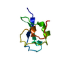
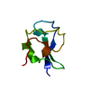
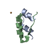
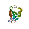
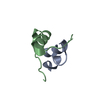
- PDB-3bbz: Structure of the nucleocapsid-binding domain from the mumps virus... -
+ Open data

- Basic information
| Entry | Database: PDB / ID: 3bbz | ||||||
|---|---|---|---|---|---|---|---|
| Title | Structure of the nucleocapsid-binding domain from the mumps virus phosphoprotein | ||||||
 Components Components | P protein | ||||||
 Keywords Keywords | VIRAL PROTEIN / REPLICATION / molten globule | ||||||
| Function / homology |  Function and homology information Function and homology information | ||||||
| Biological species |   Mumps virus Mumps virus | ||||||
| Method |  X-RAY DIFFRACTION / SYNCHROTRON / MAD / Resolution: 2.1 Å X-RAY DIFFRACTION / SYNCHROTRON / MAD / Resolution: 2.1 Å | ||||||
 Authors Authors | Kingston, R.L. / Gay, L.S. / Baase, W.S. / Matthews, B.W. | ||||||
 Citation Citation | Journal: J.Mol.Biol. / Year: 2008 Title: Structure of the nucleocapsid-binding domain from the mumps virus polymerase; an example of protein folding induced by crystallization Authors: Kingston, R.L. / Gay, L.S. / Baase, W.S. / Matthews, B.W. | ||||||
| History |
|
- Structure visualization
| Structure viewer | Molecule: MolmilJmol/JSmol |
|---|
- Downloads & links
-Download
| PDBx/mmCIF format | 3bbz.cif.gz | 31 KB | Display | PDBx/mmCIF format |
|---|---|---|---|---|
| PDB format | pdb3bbz.ent.gz | 21.6 KB | Display | PDB format |
| PDBx/mmJSON format | 3bbz.json.gz | Tree view | PDBx/mmJSON format | |
| Others |  Other downloads Other downloads |
-Validation report
| Arichive directory | https://data.pdbj.org/pub/pdb/validation_reports/bb/3bbzftp://data.pdbj.org/pub/pdb/validation_reports/bb/3bbz | HTTPS FTP |
|---|
-Related structure data
| Similar structure data |
|---|


-Links
 PDBj
PDBj


- Assembly
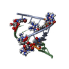
| Deposited unit | 
| ||||||||
|---|---|---|---|---|---|---|---|---|---|
| 1 | 
| ||||||||
| 2 | 
| ||||||||
| 3 | 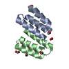
| ||||||||
| Unit cell |
|
-Components
| #1: Protein/peptide | Mass: 5455.335 Da / Num. of mol.: 2 / Fragment: nucleocapsid-binding domain, UNP residues 343-391 / Mutation: C356S Source method: isolated from a genetically manipulated source Source: (gene. exp.) Mumps virus / Strain: Jeryl-Lynn vaccine / Plasmid: pET41A(+) / Production host:  #2: Chemical |   Mass: 79.904 Da / Num. of mol.: 2 / Source method: obtained synthetically / Formula: Br Mass: 79.904 Da / Num. of mol.: 2 / Source method: obtained synthetically / Formula: Br#3: Chemical | ChemComp-FMT /   Mass: 46.025 Da / Num. of mol.: 6 / Source method: obtained synthetically / Formula: CH2O2 Mass: 46.025 Da / Num. of mol.: 6 / Source method: obtained synthetically / Formula: CH2O2#4: Water | ChemComp-HOH / |  Mass: 18.015 Da / Num. of mol.: 52 / Source method: isolated from a natural source / Formula: H2O Mass: 18.015 Da / Num. of mol.: 52 / Source method: isolated from a natural source / Formula: H2O |
|---|
-Experimental details
-Experiment
| Experiment | Method: X-RAY DIFFRACTION / Number of used crystals: 2 |
|---|
- Sample preparation
| Crystal | Density Matthews: 1.83 Å3/Da / Density % sol: 32.81 % |
|---|---|
| Crystal grow | Temperature: 277 K / Method: vapor diffusion, sitting drop / pH: 9.7 Details: 0.2M Alanine/KOH pH 9.7, 3.6-4.6M Ammonium formate, VAPOR DIFFUSION, SITTING DROP, temperature 277K |
-Data collection
| Diffraction |
| ||||||||||||||||||
|---|---|---|---|---|---|---|---|---|---|---|---|---|---|---|---|---|---|---|---|
| Diffraction source |
| ||||||||||||||||||
| Detector |
| ||||||||||||||||||
| Radiation |
| ||||||||||||||||||
| Radiation wavelength |
| ||||||||||||||||||
| Reflection | Resolution: 2.1→41.27 Å / Num. all: 4915 / Num. obs: 4915 / % possible obs: 97.2 % / Observed criterion σ(F): 0 / Observed criterion σ(I): 0 / Redundancy: 8.4 % / Biso Wilson estimate: 13 Å2 / Rmerge(I) obs: 0.14 / Rsym value: 0.14 / Net I/σ(I): 5.8 | ||||||||||||||||||
| Reflection shell | Resolution: 2.1→2.18 Å / Redundancy: 8.7 % / Rmerge(I) obs: 0.343 / Mean I/σ(I) obs: 2.4 / Num. unique all: 484 / % possible all: 98.6 |

- Processing
| Software |
| ||||||||||||||||||||||||||||||||||||||||||||||||||||||||||||||||||||||||||||||||||||||||||
|---|---|---|---|---|---|---|---|---|---|---|---|---|---|---|---|---|---|---|---|---|---|---|---|---|---|---|---|---|---|---|---|---|---|---|---|---|---|---|---|---|---|---|---|---|---|---|---|---|---|---|---|---|---|---|---|---|---|---|---|---|---|---|---|---|---|---|---|---|---|---|---|---|---|---|---|---|---|---|---|---|---|---|---|---|---|---|---|---|---|---|---|
| Refinement | Method to determine structure: MAD / Resolution: 2.1→40.74 Å / Cor.coef. Fo:Fc: 0.952 / Cor.coef. Fo:Fc free: 0.931 / SU B: 5.771 / SU ML: 0.154 / Isotropic thermal model: Isotropic / Cross valid method: THROUGHOUT / σ(F): 0 / σ(I): 0 / ESU R: 0.318 / ESU R Free: 0.219 / Stereochemistry target values: Engh & Huber
| ||||||||||||||||||||||||||||||||||||||||||||||||||||||||||||||||||||||||||||||||||||||||||
| Solvent computation | Ion probe radii: 0.8 Å / Shrinkage radii: 0.8 Å / VDW probe radii: 1.2 Å / Solvent model: MASK | ||||||||||||||||||||||||||||||||||||||||||||||||||||||||||||||||||||||||||||||||||||||||||
| Displacement parameters | Biso mean: 10.779 Å2
| ||||||||||||||||||||||||||||||||||||||||||||||||||||||||||||||||||||||||||||||||||||||||||
| Refinement step | Cycle: LAST / Resolution: 2.1→40.74 Å
| ||||||||||||||||||||||||||||||||||||||||||||||||||||||||||||||||||||||||||||||||||||||||||
| Refine LS restraints |
| ||||||||||||||||||||||||||||||||||||||||||||||||||||||||||||||||||||||||||||||||||||||||||
| LS refinement shell | Highest resolution: 2.1 Å / Num. reflection Rwork: 345 / Total num. of bins used: 20 |
