 Movie
Movie Controller

[English] 日本語
 Yorodumi
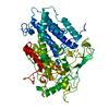
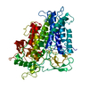
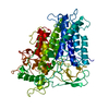
- PDB-3bb0: Crystal Structure of a Trapped Phosphate-Intermediate in Vanadium... -
+ Open data

- Basic information
| Entry | Database: PDB / ID: 3bb0 | ||||||
|---|---|---|---|---|---|---|---|
| Title | Crystal Structure of a Trapped Phosphate-Intermediate in Vanadium Apochloroperoxidase Catalyzing a Dephosphorylation Reaction | ||||||
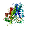
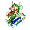
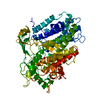
 Components Components | Vanadium chloroperoxidase | ||||||
 Keywords Keywords | OXIDOREDUCTASE / Protein phosphate-intermediate complex / phospatase activity / Chloride / Metal-binding / Peroxidase / Secreted / Vanadium | ||||||
| Function / homology |  Function and homology information Function and homology informationchloride peroxidase / chloride peroxidase activity / extracellular region / metal ion binding Similarity search - Function | ||||||
| Biological species |  Curvularia inaequalis (fungus) Curvularia inaequalis (fungus) | ||||||
| Method |  X-RAY DIFFRACTION / SYNCHROTRON / MOLECULAR REPLACEMENT / Resolution: 1.5 Å X-RAY DIFFRACTION / SYNCHROTRON / MOLECULAR REPLACEMENT / Resolution: 1.5 Å | ||||||
 Authors Authors | Messerschmidt, A. / Macedo-Ribeiro, S. | ||||||
 Citation Citation | Journal: Biochemistry / Year: 2008 Title: Crystal structure of a trapped phosphate intermediate in vanadium apochloroperoxidase catalyzing a dephosphorylation reaction Authors: Macedo-Ribeiro, S. / Renirie, R. / Wever, R. / Messerschmidt, A. #1: Journal: Proc.Natl.Acad.Sci.Usa / Year: 1996Title: X-ray structure of a vanadium-containing enzyme: chloroperoxidase from the fungus Curvularia inaequalis Authors: Messerschmidt, A. / Wever, R. #2: Journal: J.Biol.Inorg.Chem. / Year: 1999Title: X-ray crystal structures of active site mutants of the vanadium-containing chloroperoxidase from the fungus Curvularia inaequalis Authors: Macedo-Ribeiro, S. / Hemrika, W. / Renirie, R. / Wever, R. / Messerschmidt, A. | ||||||
| History |
|
- Structure visualization
| Structure viewer | Molecule: MolmilJmol/JSmol |
|---|
- Downloads & links
-Download
| PDBx/mmCIF format | 3bb0.cif.gz | 144.8 KB | Display | PDBx/mmCIF format |
|---|---|---|---|---|
| PDB format | pdb3bb0.ent.gz | 111.2 KB | Display | PDB format |
| PDBx/mmJSON format | 3bb0.json.gz | Tree view | PDBx/mmJSON format | |
| Others |  Other downloads Other downloads |
-Validation report
| Arichive directory | https://data.pdbj.org/pub/pdb/validation_reports/bb/3bb0ftp://data.pdbj.org/pub/pdb/validation_reports/bb/3bb0 | HTTPS FTP |
|---|
-Related structure data
| Related structure data | 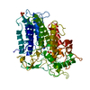 1vnsS S: Starting model for refinement |
|---|---|
| Similar structure data |


-Links
 PDBj
PDBj- Assembly
| Deposited unit | 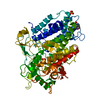
| ||||||||||||
|---|---|---|---|---|---|---|---|---|---|---|---|---|---|
| 1 |
| ||||||||||||
| Unit cell |
| ||||||||||||
| Components on special symmetry positions |
|
-Components
| #1: Protein | Mass: 67671.523 Da / Num. of mol.: 1 Source method: isolated from a genetically manipulated source Source: (gene. exp.) Curvularia inaequalis (fungus) / Gene: CPO / Plasmid: pTNT14 / Production host:  | ||||||
|---|---|---|---|---|---|---|---|
| #2: Chemical | ChemComp-PO3 /   Mass: 78.972 Da / Num. of mol.: 1 / Source method: obtained synthetically / Formula: PO3 Mass: 78.972 Da / Num. of mol.: 1 / Source method: obtained synthetically / Formula: PO3 | ||||||
| #3: Chemical |   Mass: 96.063 Da / Num. of mol.: 2 / Source method: obtained synthetically / Formula: SO4 Mass: 96.063 Da / Num. of mol.: 2 / Source method: obtained synthetically / Formula: SO4#4: Water | ChemComp-HOH / |  Mass: 18.015 Da / Num. of mol.: 795 / Source method: isolated from a natural source / Formula: H2O Mass: 18.015 Da / Num. of mol.: 795 / Source method: isolated from a natural source / Formula: H2OHas protein modification | Y | Sequence details | THIS CONFLICT MAY BE A SEQUENCING ERROR (THE CODON FOR ARG VARIES BY ONE NUCLEOTIDE CHANGE WITH PRO ...THIS CONFLICT MAY BE A SEQUENCING | |
-Experimental details
-Experiment
| Experiment | Method: X-RAY DIFFRACTION / Number of used crystals: 1 |
|---|
- Sample preparation
| Crystal | Density Matthews: 2.41 Å3/Da / Density % sol: 48.94 % |
|---|---|
| Crystal grow | Temperature: 277 K / Method: vapor diffusion, hanging drop / pH: 8 Details: 3 micro-l of protein solution (5mg/ml in 5mM MOPS, pH 7.5), 3 micro-l precipitating buffer (1.7M ammonium sulfate, 0.1M Tris-HCl, pH 8.0, 0.3 micro-l 10mM cysteine-HCl) , VAPOR DIFFUSION, ...Details: 3 micro-l of protein solution (5mg/ml in 5mM MOPS, pH 7.5), 3 micro-l precipitating buffer (1.7M ammonium sulfate, 0.1M Tris-HCl, pH 8.0, 0.3 micro-l 10mM cysteine-HCl) , VAPOR DIFFUSION, HANGING DROP, temperature 277K |
-Data collection
| Diffraction | Mean temperature: 100 K |
|---|---|
| Diffraction source | Source: SYNCHROTRON / Site: MPG/DESY, HAMBURG  / Beamline: BW6 / Wavelength: 1.05 Å / Beamline: BW6 / Wavelength: 1.05 Å |
| Detector | Type: MAR scanner 345 mm plate / Detector: IMAGE PLATE / Date: Jun 14, 1998 / Details: mirrors |
| Radiation | Monochromator: Mirrors / Protocol: SINGLE WAVELENGTH / Monochromatic (M) / Laue (L): M / Scattering type: x-ray |
| Radiation wavelength | Wavelength: 1.05 Å / Relative weight: 1 |
| Reflection | Resolution: 1.5→50 Å / Num. all: 101188 / Num. obs: 100278 / % possible obs: 99.1 % / Observed criterion σ(F): 2 / Observed criterion σ(I): 2 / Redundancy: 1.8 % / Rmerge(I) obs: 0.062 / Rsym value: 0.062 / Net I/σ(I): 8.2 |
| Reflection shell | Resolution: 1.5→1.53 Å / Redundancy: 1.8 % / Rmerge(I) obs: 0.317 / Mean I/σ(I) obs: 3.5 / Num. unique all: 5046 / Rsym value: 0.317 / % possible all: 96 |
- Processing
| Software |
| ||||||||||||||||||||||||||||||||||||||||||||||||||||||||||||||||||||||||||||||||||||||||||
|---|---|---|---|---|---|---|---|---|---|---|---|---|---|---|---|---|---|---|---|---|---|---|---|---|---|---|---|---|---|---|---|---|---|---|---|---|---|---|---|---|---|---|---|---|---|---|---|---|---|---|---|---|---|---|---|---|---|---|---|---|---|---|---|---|---|---|---|---|---|---|---|---|---|---|---|---|---|---|---|---|---|---|---|---|---|---|---|---|---|---|---|
| Refinement | Method to determine structure: MOLECULAR REPLACEMENT Starting model: PDB ENTRY 1VNS Resolution: 1.5→14.9 Å / Cor.coef. Fo:Fc: 0.964 / Cor.coef. Fo:Fc free: 0.955 / SU B: 1.066 / SU ML: 0.041 / Isotropic thermal model: isotropic / Cross valid method: THROUGHOUT / σ(F): 0 / σ(I): 0 / ESU R: 0.071 / ESU R Free: 0.07 / Stereochemistry target values: MAXIMUM LIKELIHOOD / Details: HYDROGENS HAVE BEEN ADDED IN THE RIDING POSITIONS
| ||||||||||||||||||||||||||||||||||||||||||||||||||||||||||||||||||||||||||||||||||||||||||
| Solvent computation | Ion probe radii: 0.8 Å / Shrinkage radii: 0.8 Å / VDW probe radii: 1.4 Å / Solvent model: BABINET MODEL WITH MASK | ||||||||||||||||||||||||||||||||||||||||||||||||||||||||||||||||||||||||||||||||||||||||||
| Displacement parameters | Biso mean: 17.863 Å2
| ||||||||||||||||||||||||||||||||||||||||||||||||||||||||||||||||||||||||||||||||||||||||||
| Refine analyze |
| ||||||||||||||||||||||||||||||||||||||||||||||||||||||||||||||||||||||||||||||||||||||||||
| Refinement step | Cycle: LAST / Resolution: 1.5→14.9 Å
| ||||||||||||||||||||||||||||||||||||||||||||||||||||||||||||||||||||||||||||||||||||||||||
| Refine LS restraints |
| ||||||||||||||||||||||||||||||||||||||||||||||||||||||||||||||||||||||||||||||||||||||||||
| LS refinement shell | Resolution: 1.5→1.539 Å / Total num. of bins used: 20
|
