 Movie
Movie Controller

[English] 日本語
 Yorodumi
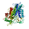
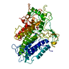
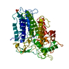
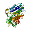
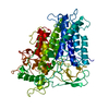
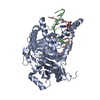
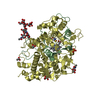
- PDB-1idu: CRYSTAL STRUCTURE OF THE PEROXIDE FORM OF THE VANADIUM-CONTAINING... -
+ Open data

- Basic information
| Entry | Database: PDB / ID: 1idu | ||||||
|---|---|---|---|---|---|---|---|
| Title | CRYSTAL STRUCTURE OF THE PEROXIDE FORM OF THE VANADIUM-CONTAINING CHLOROPEROXIDASE FROM CURVULARIA INAEQUALIS | ||||||
 Components Components | VANADIUM CHLOROPEROXIDASE | ||||||
 Keywords Keywords | OXIDOREDUCTASE / Two four-helix bundles / peroxide derivative | ||||||
| Function / homology |  Function and homology information Function and homology informationchloride peroxidase / chloride peroxidase activity / extracellular region / metal ion binding Similarity search - Function | ||||||
| Biological species |  Curvularia inaequalis (fungus) Curvularia inaequalis (fungus) | ||||||
| Method |  X-RAY DIFFRACTION / FOURIER SYNTHESIS / Resolution: 2.24 Å X-RAY DIFFRACTION / FOURIER SYNTHESIS / Resolution: 2.24 Å | ||||||
 Authors Authors | Messerschmidt, A. / Prade, L. / Wever, R. | ||||||
 Citation Citation | Journal: Biol.Chem. / Year: 1997 Title: Implications for the catalytic mechanism of the vanadium-containing enzyme chloroperoxidase from the fungus Curvularia inaequalis by X-ray structures of the native and peroxide form. Authors: Messerschmidt, A. / Prade, L. / Wever, R. | ||||||
| History |
|
- Structure visualization
| Structure viewer | Molecule: MolmilJmol/JSmol |
|---|
- Downloads & links
-Download
| PDBx/mmCIF format | 1idu.cif.gz | 139.9 KB | Display | PDBx/mmCIF format |
|---|---|---|---|---|
| PDB format | pdb1idu.ent.gz | 105.4 KB | Display | PDB format |
| PDBx/mmJSON format | 1idu.json.gz | Tree view | PDBx/mmJSON format | |
| Others |  Other downloads Other downloads |
-Validation report
| Arichive directory | https://data.pdbj.org/pub/pdb/validation_reports/id/1iduftp://data.pdbj.org/pub/pdb/validation_reports/id/1idu | HTTPS FTP |
|---|
-Related structure data
| Related structure data | 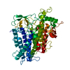 1idqC 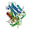 1vncS C: citing same article ( S: Starting model for refinement |
|---|---|
| Similar structure data |


-Links
 PDBj
PDBj- Assembly
| Deposited unit | 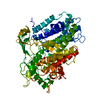
| |||||||||
|---|---|---|---|---|---|---|---|---|---|---|
| 1 |
| |||||||||
| Unit cell |
| |||||||||
| Components on special symmetry positions |
| |||||||||
| Details | The biologically active molecule is a monomer. |
-Components
| #1: Protein | Mass: 67603.398 Da / Num. of mol.: 1 / Source method: isolated from a natural source / Source: (natural) Curvularia inaequalis (fungus)Strain: STRAIN 102.42 FROM CENTRAAL BUREAU VOR SCHIMMELCULTURES (CBS, BAARN, THE NETHERLANDS) References: UniProt: P49053, chloride peroxidase |
|---|---|
| #2: Chemical | ChemComp-VO4 /   Mass: 114.939 Da / Num. of mol.: 1 / Source method: obtained synthetically / Formula: VO4 Mass: 114.939 Da / Num. of mol.: 1 / Source method: obtained synthetically / Formula: VO4 |
| #3: Water | ChemComp-HOH /  Mass: 18.015 Da / Num. of mol.: 604 / Source method: isolated from a natural source / Formula: H2O Mass: 18.015 Da / Num. of mol.: 604 / Source method: isolated from a natural source / Formula: H2O |
-Experimental details
-Experiment
| Experiment | Method: X-RAY DIFFRACTION / Number of used crystals: 1 |
|---|
- Sample preparation
| Crystal | Density Matthews: 2.68 Å3/Da / Density % sol: 54.15 % | ||||||||||||||||||||||||||||||||||||||||||||||||||||||||||||
|---|---|---|---|---|---|---|---|---|---|---|---|---|---|---|---|---|---|---|---|---|---|---|---|---|---|---|---|---|---|---|---|---|---|---|---|---|---|---|---|---|---|---|---|---|---|---|---|---|---|---|---|---|---|---|---|---|---|---|---|---|---|
| Crystal grow | Temperature: 294 K / Method: vapor diffusion, hanging drop / pH: 8 Details: Reservoir: 1.5 M ammonium sulfate, 0.1 M Tris-HCl; protein drop: 7.5 microliter protein solution (8.7 mg/ml), 1 mM Na3VO4 plus 2.5 microliter reservoir solution. The peroxide complex was ...Details: Reservoir: 1.5 M ammonium sulfate, 0.1 M Tris-HCl; protein drop: 7.5 microliter protein solution (8.7 mg/ml), 1 mM Na3VO4 plus 2.5 microliter reservoir solution. The peroxide complex was produced by soaking the crystals in mother liquor (2M ammonium sulfate, 0.1 M Tris-H2SO4, pH 8.0), containing 20 mM peroxide, for two hours and then flash cooled., VAPOR DIFFUSION, HANGING DROP, temperature 294K | ||||||||||||||||||||||||||||||||||||||||||||||||||||||||||||
| Crystal | *PLUS Density % sol: 55 % | ||||||||||||||||||||||||||||||||||||||||||||||||||||||||||||
| Crystal grow | *PLUS Temperature: 21 ℃ | ||||||||||||||||||||||||||||||||||||||||||||||||||||||||||||
| Components of the solutions | *PLUS
|
-Data collection
| Diffraction | Mean temperature: 100 K |
|---|---|
| Diffraction source | Source: ROTATING ANODE / Type: RIGAKU RU200 / Wavelength: 1.5418 Å |
| Detector | Type: MARRESEARCH / Detector: IMAGE PLATE / Date: Jun 23, 1996 / Details: Graphite monochromator |
| Radiation | Monochromator: Graphite / Protocol: SINGLE WAVELENGTH / Monochromatic (M) / Laue (L): M / Scattering type: x-ray |
| Radiation wavelength | Wavelength: 1.5418 Å / Relative weight: 1 |
| Reflection | Resolution: 2.24→33.84 Å / Num. all: 33853 / Num. obs: 29878 / % possible obs: 88.3 % / Observed criterion σ(F): 0 / Observed criterion σ(I): 0 / Redundancy: 2.1 % / Biso Wilson estimate: 34 Å2 / Rmerge(I) obs: 0.118 / Rsym value: 0.114 / Net I/σ(I): 7.5 |
| Reflection shell | Resolution: 2.24→2.34 Å / Redundancy: 2 % / Rmerge(I) obs: 0.297 / Mean I/σ(I) obs: 1.2 / Num. unique all: 3750 / Rsym value: 0.293 / % possible all: 40.2 |
| Reflection | *PLUS Num. measured all: 102166 |
| Reflection shell | *PLUS % possible obs: 40.2 % |
- Processing
| Software |
| ||||||||||||||||||||||||||||||||||||
|---|---|---|---|---|---|---|---|---|---|---|---|---|---|---|---|---|---|---|---|---|---|---|---|---|---|---|---|---|---|---|---|---|---|---|---|---|---|
| Refinement | Method to determine structure: FOURIER SYNTHESIS Starting model: PDB ENTRY 1VNC Resolution: 2.24→8 Å / Isotropic thermal model: Isotropic / σ(F): 0 / σ(I): 0 / Stereochemistry target values: Engh & Huber
| ||||||||||||||||||||||||||||||||||||
| Displacement parameters | Biso mean: 29.13 Å2 | ||||||||||||||||||||||||||||||||||||
| Refine analyze | Luzzati coordinate error obs: 0.23 Å / Luzzati d res low obs: 8 Å | ||||||||||||||||||||||||||||||||||||
| Refinement step | Cycle: LAST / Resolution: 2.24→8 Å
| ||||||||||||||||||||||||||||||||||||
| Refine LS restraints |
| ||||||||||||||||||||||||||||||||||||
| LS refinement shell |
| ||||||||||||||||||||||||||||||||||||
| Software | *PLUS Name: X-PLOR / Version: 3.843 / Classification: refinement | ||||||||||||||||||||||||||||||||||||
| Refine LS restraints | *PLUS
|
