- PDB-3b5d: EmrE multidrug transporter in complex with TPP, C2 crystal form -
+
Open data
ID or keywords:
Loading...
-
Basic information
Entry
Database: PDB / ID: 3b5d
Title
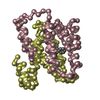
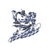
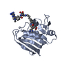
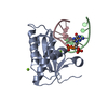
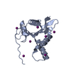
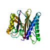
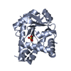
EmrE multidrug transporter in complex with TPP, C2 crystal form
Components
Multidrug transporter emrE
Keywords
MEMBRANE PROTEIN / HELICAL MEMBRANE PROTEIN / MULTIDRUG RESISTANCE TRANSPORTER / SMR / Antiport / Inner membrane / Transmembrane / Joint Center for Innovative Membrane Protein Technologies
Function / homology
Function and homology information
EmrE multidrug transporter complex / amino-acid betaine transmembrane transporter activity / glycine betaine transport / choline transmembrane transporter activity / choline transport / xenobiotic detoxification by transmembrane export across the plasma membrane / antiporter activity / response to osmotic stress / Antimicrobial resistance / xenobiotic transmembrane transporter activity ...EmrE multidrug transporter complex / amino-acid betaine transmembrane transporter activity / glycine betaine transport / choline transmembrane transporter activity / choline transport / xenobiotic detoxification by transmembrane export across the plasma membrane / antiporter activity / response to osmotic stress / Antimicrobial resistance / xenobiotic transmembrane transporter activity / transmembrane transporter activity / xenobiotic transport / xenobiotic metabolic process / transmembrane transport / cellular response to xenobiotic stimulus / response to xenobiotic stimulus / DNA damage response / membrane / identical protein binding / plasma membrane Similarity search - Function
Small drug/metabolite transporter protein family / Small multidrug resistance protein / Small Multidrug Resistance protein / : Similarity search - Domain/homology
MultidrugtransporteremrE / Efflux-multidrug resistance protein emrE / Methyl viologen resistance protein C / Ethidium resistance protein / Coordinate model: Cα atoms only
Mass: 11963.278 Da / Num. of mol.: 2 Source method: isolated from a genetically manipulated source Source: (gene. exp.) Escherichia coli K12 (bacteria) / Species: Escherichia coli / Strain: K-12 / Gene: emrE, eb, mvrC / Plasmid: pIVEX / Species (production host): Escherichia coli / Production host: Escherichia coli BL21(DE3) (bacteria) / Strain (production host): BL21(DE3) / References: UniProt: P23895
In the structure databanks used in Yorodumi, some data are registered as the other names, "COVID-19 virus" and "2019-nCoV". Here are the details of the virus and the list of structure data.
Jan 31, 2019. EMDB accession codes are about to change! (news from PDBe EMDB page)
EMDB accession codes are about to change! (news from PDBe EMDB page)
The allocation of 4 digits for EMDB accession codes will soon come to an end. Whilst these codes will remain in use, new EMDB accession codes will include an additional digit and will expand incrementally as the available range of codes is exhausted. The current 4-digit format prefixed with “EMD-” (i.e. EMD-XXXX) will advance to a 5-digit format (i.e. EMD-XXXXX), and so on. It is currently estimated that the 4-digit codes will be depleted around Spring 2019, at which point the 5-digit format will come into force.
The EM Navigator/Yorodumi systems omit the EMD- prefix.
Related info.:Q: What is EMD? / ID/Accession-code notation in Yorodumi/EM Navigator
Yorodumi is a browser for structure data from EMDB, PDB, SASBDB, etc.
This page is also the successor to EM Navigator detail page, and also detail information page/front-end page for Omokage search.
The word "yorodu" (or yorozu) is an old Japanese word meaning "ten thousand". "mi" (miru) is to see.
Related info.:EMDB / PDB / SASBDB / Comparison of 3 databanks / Yorodumi Search / Aug 31, 2016. New EM Navigator & Yorodumi / Yorodumi Papers / Jmol/JSmol / Function and homology information / Changes in new EM Navigator and Yorodumi
 Movie
Movie Controller


 Open data
 Basic information
 Components
 Keywords
 Function and homology information
 X-RAY DIFFRACTION /
  Authors
 Citation
 Structure visualization
 Downloads & links
 Other downloads
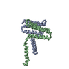


 PDBj
PDBj
 Assembly
 Mass: 339.389 Da / Num. of mol.: 1 / Source method: obtained synthetically / Formula: C24H20P
 Sample preparation
 / Beamline: BL11-1 / Wavelength: 0.9790, 0.9793, 0.9184
 Processing