- PDB-6tuu: Leishmania infantum Rad51 surrogate LiRadA10 in complex with 5,6,... -
+
Open data
ID or keywords:
Loading...
-
Basic information
Entry
Database: PDB / ID: 6tuu
Title
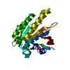
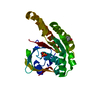
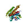
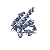
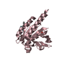
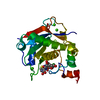
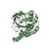
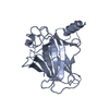
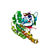
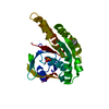
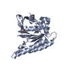
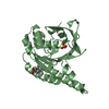
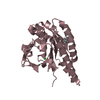
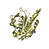
Leishmania infantum Rad51 surrogate LiRadA10 in complex with 5,6,7,8-tetrahydro-2-naphthoic acid
Components
DNA repair and recombination protein RadA
Keywords
RECOMBINATION / Recombinase / FBDD / Fragment / Surrogate / RadA / Rad51 / Archaeal
Function / homology
Function and homology information
ATP-dependent DNA damage sensor activity / DNA recombination / damaged DNA binding / DNA repair / ATP binding Similarity search - Function
DNA recombination/repair protein RadA / DNA recombination and repair protein, RecA-like / DNA recombination and repair protein Rad51-like, C-terminal / Rad51 / DNA recombination and repair protein RecA, monomer-monomer interface / RecA family profile 2. / DNA recombination and repair protein RecA-like, ATP-binding domain / RecA family profile 1. / DNA repair Rad51/transcription factor NusA, alpha-helical / SAM-like Helix-hairpin-helix tandem ...DNA recombination/repair protein RadA / DNA recombination and repair protein, RecA-like / DNA recombination and repair protein Rad51-like, C-terminal / Rad51 / DNA recombination and repair protein RecA, monomer-monomer interface / RecA family profile 2. / DNA recombination and repair protein RecA-like, ATP-binding domain / RecA family profile 1. / DNA repair Rad51/transcription factor NusA, alpha-helical / SAM-like Helix-hairpin-helix tandem / Helix-hairpin-helix DNA-binding motif, class 1 / Helix-hairpin-helix DNA-binding motif class 1 / ATPases associated with a variety of cellular activities / AAA+ ATPase domain / P-loop containing nucleoside triphosphate hydrolase Similarity search - Domain/homology
5,6,7,8-tetrahydronaphthalene-2-carboxylic acid / PHOSPHATE ION / DNA repair and recombination protein RadA Similarity search - Component
A: DNA repair and recombination protein RadA B: DNA repair and recombination protein RadA C: DNA repair and recombination protein RadA D: DNA repair and recombination protein RadA hetero molecules
In the structure databanks used in Yorodumi, some data are registered as the other names, "COVID-19 virus" and "2019-nCoV". Here are the details of the virus and the list of structure data.
Jan 31, 2019. EMDB accession codes are about to change! (news from PDBe EMDB page)
EMDB accession codes are about to change! (news from PDBe EMDB page)
The allocation of 4 digits for EMDB accession codes will soon come to an end. Whilst these codes will remain in use, new EMDB accession codes will include an additional digit and will expand incrementally as the available range of codes is exhausted. The current 4-digit format prefixed with “EMD-” (i.e. EMD-XXXX) will advance to a 5-digit format (i.e. EMD-XXXXX), and so on. It is currently estimated that the 4-digit codes will be depleted around Spring 2019, at which point the 5-digit format will come into force.
The EM Navigator/Yorodumi systems omit the EMD- prefix.
Related info.:Q: What is EMD? / ID/Accession-code notation in Yorodumi/EM Navigator
Yorodumi is a browser for structure data from EMDB, PDB, SASBDB, etc.
This page is also the successor to EM Navigator detail page, and also detail information page/front-end page for Omokage search.
The word "yorodu" (or yorozu) is an old Japanese word meaning "ten thousand". "mi" (miru) is to see.
Related info.:EMDB / PDB / SASBDB / Comparison of 3 databanks / Yorodumi Search / Aug 31, 2016. New EM Navigator & Yorodumi / Yorodumi Papers / Jmol/JSmol / Function and homology information / Changes in new EM Navigator and Yorodumi
 Movie
Movie Controller

 Yorodumi
 Open data
 Basic information
 Components
 Keywords
 Function and homology information
 Pyrococcus furiosus (archaea)
 X-RAY DIFFRACTION /
  Authors
 United Kingdom, 1items
  Citation
 Structure visualization
 Downloads & links
 Other downloads
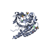
 PDBj
PDBj


 Assembly
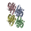


 Mass: 94.971 Da / Num. of mol.: 2 / Source method: obtained synthetically / Formula: PO4
 Mass: 176.212 Da / Num. of mol.: 4 / Source method: obtained synthetically / Formula: C11H12O2 / Feature type: SUBJECT OF INVESTIGATION
 Mass: 35.453 Da / Num. of mol.: 1 / Source method: isolated from a natural source / Formula: Cl
 Mass: 18.015 Da / Num. of mol.: 600 / Source method: isolated from a natural source / Formula: H2O
 Sample preparation
 Processing