 Movie
Movie Controller

[English] 日本語
 Yorodumi
- PDB-3axy: Structure of Florigen Activation Complex Consisting of Rice Flori... -
+ Open data

- Basic information
| Entry | Database: PDB / ID: 3axy | ||||||
|---|---|---|---|---|---|---|---|
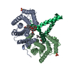
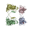
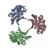
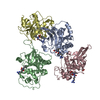
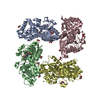
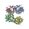
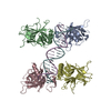
| Title | Structure of Florigen Activation Complex Consisting of Rice Florigen Hd3a, 14-3-3 Protein GF14 and Rice FD Homolog OsFD1 | ||||||
 Components Components |
| ||||||
 Keywords Keywords | SIGNALING PROTEIN/PROTEIN BINDING / Phosphatidylethanolamine-binding Protein / 14-3-3 Protein / bZip protein / Floral Induction / Transcriptional Activator / Signaling Protein / DNA Binding / Phosphorylation / Nucleus / SIGNALING PROTEIN-PROTEIN BINDING complex | ||||||
| Function / homology |  Function and homology information Function and homology informationshort-day photoperiodism, flowering / regulation of timing of transition from vegetative to reproductive phase / short-day photoperiodism / regulation of flower development / vegetative to reproductive phase transition of meristem / inflorescence development / flower development / phosphatidylethanolamine binding / intracellular protein localization / cell differentiation ...short-day photoperiodism, flowering / regulation of timing of transition from vegetative to reproductive phase / short-day photoperiodism / regulation of flower development / vegetative to reproductive phase transition of meristem / inflorescence development / flower development / phosphatidylethanolamine binding / intracellular protein localization / cell differentiation / signal transduction / nucleus / cytoplasm Similarity search - Function | ||||||
| Biological species |  | ||||||
| Method |  X-RAY DIFFRACTION / SYNCHROTRON / MOLECULAR REPLACEMENT / Resolution: 2.4 Å X-RAY DIFFRACTION / SYNCHROTRON / MOLECULAR REPLACEMENT / Resolution: 2.4 Å | ||||||
 Authors Authors | Ohki, I. / Furuita, K. / Hayashi, K. / Taoka, K. / Tsuji, H. / Nakagawa, A. / Shimamoto, K. / Kojima, C. | ||||||
 Citation Citation | Journal: Nature / Year: 2011 Title: 14-3-3 proteins act as intracellular receptors for rice Hd3a florigen Authors: Taoka, K. / Ohki, I. / Tsuji, H. / Furuita, K. / Hayashi, K. / Yanase, T. / Yamaguchi, M. / Nakashima, C. / Purwestri, Y.A. / Tamaki, S. / Ogaki, Y. / Shimada, C. / Nakagawa, A. / Kojima, C. / Shimamoto, K. | ||||||
| History |
|
- Structure visualization
| Structure viewer | Molecule: MolmilJmol/JSmol |
|---|
- Downloads & links
-Download
| PDBx/mmCIF format | 3axy.cif.gz | 553.5 KB | Display | PDBx/mmCIF format |
|---|---|---|---|---|
| PDB format | pdb3axy.ent.gz | 457 KB | Display | PDB format |
| PDBx/mmJSON format | 3axy.json.gz | Tree view | PDBx/mmJSON format | |
| Others |  Other downloads Other downloads |
-Validation report
| Arichive directory | https://data.pdbj.org/pub/pdb/validation_reports/ax/3axyftp://data.pdbj.org/pub/pdb/validation_reports/ax/3axy | HTTPS FTP |
|---|
-Related structure data

-Links
 PDBj
PDBj
- Assembly
| Deposited unit | 
| ||||||||
|---|---|---|---|---|---|---|---|---|---|
| 1 | 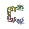
| ||||||||
| 2 | 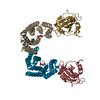
| ||||||||
| Unit cell |
|
-Components
| #1: Protein | Mass: 18982.418 Da / Num. of mol.: 4 / Fragment: UNP residues 6-170 / Mutation: C43L,C109S,C166S Source method: isolated from a genetically manipulated source Source: (gene. exp.) Gene: HD3A, LOC_Os06g06320, Os06g0157700, OsJ_20191, P0046E09.30, P0702F05.10 Plasmid: pCOLD / Production host:  #2: Protein | Mass: 27151.787 Da / Num. of mol.: 4 / Fragment: UNP residues 1-235 Source method: isolated from a genetically manipulated source Source: (gene. exp.) Gene: GF14C, LOC_Os08g33370, OJ1124_B05.7, Os08g0430500 / Plasmid: pGEX6P3 / Production host: #3: Protein/peptide | Mass: 1111.207 Da / Num. of mol.: 4 / Fragment: C-TERMINAL MOTIF / Source method: obtained synthetically / Details: OsFD1(187-195) fragment, phosphorylated at S192 #4: Water | ChemComp-HOH / |  Mass: 18.015 Da / Num. of mol.: 777 / Source method: isolated from a natural source / Formula: H2O Mass: 18.015 Da / Num. of mol.: 777 / Source method: isolated from a natural source / Formula: H2OHas protein modification | Y | |
|---|
-Experimental details
-Experiment
| Experiment | Method: X-RAY DIFFRACTION / Number of used crystals: 1 |
|---|
- Sample preparation
| Crystal | Density Matthews: 3.54 Å3/Da / Density % sol: 65.28 % |
|---|---|
| Crystal grow | Temperature: 277 K / Method: vapor diffusion, sitting drop / pH: 7.5 Details: 0.1M HEPES (pH 7.5), 0.2M ammonium sulfate, 25% PEG 3350, VAPOR DIFFUSION, SITTING DROP, temperature 277K |
-Data collection
| Diffraction | Mean temperature: 100 K |
|---|---|
| Diffraction source | Source: SYNCHROTRON / Site: Photon Factory  / Beamline: BL-5A / Wavelength: 1 Å / Beamline: BL-5A / Wavelength: 1 Å |
| Detector | Type: ADSC QUANTUM 315 / Detector: CCD / Date: Jun 5, 2009 |
| Radiation | Monochromator: Si(111) double crystal monochromator / Protocol: SINGLE WAVELENGTH / Monochromatic (M) / Laue (L): M / Scattering type: x-ray |
| Radiation wavelength | Wavelength: 1 Å / Relative weight: 1 |
| Reflection | Resolution: 2.4→50 Å / Num. obs: 101603 / % possible obs: 98.1 % / Observed criterion σ(I): 3 / Redundancy: 2.7 % / Rsym value: 0.067 / Net I/σ(I): 12.4 |
| Reflection shell | Resolution: 2.4→2.49 Å / Redundancy: 2.7 % / Mean I/σ(I) obs: 3.3 / Rsym value: 0.252 / % possible all: 97.1 |
- Processing
| Software |
| |||||||||||||||||||||||||||||||||||||||||||||||||||||||||||||||||||||||||||||||||||||||||||||||||||||||||||||||||||||||||||||||||||||||||||||||||||||||||||||||||||||||||||||||
|---|---|---|---|---|---|---|---|---|---|---|---|---|---|---|---|---|---|---|---|---|---|---|---|---|---|---|---|---|---|---|---|---|---|---|---|---|---|---|---|---|---|---|---|---|---|---|---|---|---|---|---|---|---|---|---|---|---|---|---|---|---|---|---|---|---|---|---|---|---|---|---|---|---|---|---|---|---|---|---|---|---|---|---|---|---|---|---|---|---|---|---|---|---|---|---|---|---|---|---|---|---|---|---|---|---|---|---|---|---|---|---|---|---|---|---|---|---|---|---|---|---|---|---|---|---|---|---|---|---|---|---|---|---|---|---|---|---|---|---|---|---|---|---|---|---|---|---|---|---|---|---|---|---|---|---|---|---|---|---|---|---|---|---|---|---|---|---|---|---|---|---|---|---|---|---|---|
| Refinement | Method to determine structure: MOLECULAR REPLACEMENT Starting model: PDB Code 1WKP for Hd3a, 2O98 for GF14c Resolution: 2.4→50 Å / Cor.coef. Fo:Fc: 0.917 / Cor.coef. Fo:Fc free: 0.878 / SU B: 12.325 / SU ML: 0.188 / Cross valid method: THROUGHOUT / ESU R: 0.342 / ESU R Free: 0.264 / Stereochemistry target values: MAXIMUM LIKELIHOOD / Details: HYDROGENS HAVE BEEN ADDED IN THE RIDING POSITIONS
| |||||||||||||||||||||||||||||||||||||||||||||||||||||||||||||||||||||||||||||||||||||||||||||||||||||||||||||||||||||||||||||||||||||||||||||||||||||||||||||||||||||||||||||||
| Solvent computation | Ion probe radii: 0.8 Å / Shrinkage radii: 0.8 Å / VDW probe radii: 1.4 Å / Solvent model: MASK | |||||||||||||||||||||||||||||||||||||||||||||||||||||||||||||||||||||||||||||||||||||||||||||||||||||||||||||||||||||||||||||||||||||||||||||||||||||||||||||||||||||||||||||||
| Displacement parameters | Biso mean: 32.061 Å2
| |||||||||||||||||||||||||||||||||||||||||||||||||||||||||||||||||||||||||||||||||||||||||||||||||||||||||||||||||||||||||||||||||||||||||||||||||||||||||||||||||||||||||||||||
| Refinement step | Cycle: LAST / Resolution: 2.4→50 Å
| |||||||||||||||||||||||||||||||||||||||||||||||||||||||||||||||||||||||||||||||||||||||||||||||||||||||||||||||||||||||||||||||||||||||||||||||||||||||||||||||||||||||||||||||
| Refine LS restraints |
| |||||||||||||||||||||||||||||||||||||||||||||||||||||||||||||||||||||||||||||||||||||||||||||||||||||||||||||||||||||||||||||||||||||||||||||||||||||||||||||||||||||||||||||||
| LS refinement shell | Resolution: 2.4→2.462 Å / Total num. of bins used: 20
| |||||||||||||||||||||||||||||||||||||||||||||||||||||||||||||||||||||||||||||||||||||||||||||||||||||||||||||||||||||||||||||||||||||||||||||||||||||||||||||||||||||||||||||||
| Refinement TLS params. | Method: refined / Refine-ID: X-RAY DIFFRACTION
| |||||||||||||||||||||||||||||||||||||||||||||||||||||||||||||||||||||||||||||||||||||||||||||||||||||||||||||||||||||||||||||||||||||||||||||||||||||||||||||||||||||||||||||||
| Refinement TLS group |
|
