| Entry | Database: PDB / ID: 3axl
|
|---|
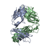
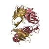
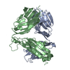
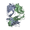
| Title | Murine Valpha 10 Vbeta 8.1 T-cell receptor |
|---|
 Components Components | |
|---|
 Keywords Keywords | IMMUNE SYSTEM / Immunoglobulin fold / T-cell receptor / CD1d binding |
|---|
| Function / homology | Immunoglobulins / Immunoglobulin-like / Sandwich / Mainly Beta Function and homology information Function and homology information |
|---|
| Biological species |   Mus musculus (house mouse) Mus musculus (house mouse) |
|---|
| Method |  X-RAY DIFFRACTION / SYNCHROTRON / MOLECULAR REPLACEMENT / Resolution: 2.9 Å X-RAY DIFFRACTION / SYNCHROTRON / MOLECULAR REPLACEMENT / Resolution: 2.9 Å |
|---|
 Authors Authors | Patel, O. / Rossjohn, J. |
|---|
 Citation Citation | Journal: Nat.Immunol. / Year: 2011
Title: A semi-invariant V(alpha)10(+) T cell antigen receptor defines a population of natural killer T cells with distinct glycolipid antigen-recognition properties
Authors: Uldrich, A.P. / Patel, O. / Cameron, G. / Pellicci, D.G. / Day, E.B. / Sullivan, L.C. / Kyparissoudis, K. / Kjer-Nielsen, L. / Vivian, J.P. / Cao, B. / Brooks, A.G. / Williams, S.J. / ...Authors: Uldrich, A.P. / Patel, O. / Cameron, G. / Pellicci, D.G. / Day, E.B. / Sullivan, L.C. / Kyparissoudis, K. / Kjer-Nielsen, L. / Vivian, J.P. / Cao, B. / Brooks, A.G. / Williams, S.J. / Illarionov, P. / Besra, G.S. / Turner, S.J. / Porcelli, S.A. / McCluskey, J. / Smyth, M.J. / Rossjohn, J. / Godfrey, D.I. |
|---|
| History | | Deposition | Apr 11, 2011 | Deposition site: PDBJ / Processing site: PDBJ |
|---|
| Revision 1.0 | Aug 3, 2011 | Provider: repository / Type: Initial release |
|---|
| Revision 1.1 | Oct 9, 2024 | Group: Data collection / Database references ...Data collection / Database references / Refinement description / Structure summary
Category: chem_comp_atom / chem_comp_bond ...chem_comp_atom / chem_comp_bond / database_2 / pdbx_entry_details / pdbx_modification_feature / struct_ncs_dom_lim
Item: _database_2.pdbx_DOI / _database_2.pdbx_database_accession ..._database_2.pdbx_DOI / _database_2.pdbx_database_accession / _struct_ncs_dom_lim.beg_auth_comp_id / _struct_ncs_dom_lim.beg_label_asym_id / _struct_ncs_dom_lim.beg_label_comp_id / _struct_ncs_dom_lim.beg_label_seq_id / _struct_ncs_dom_lim.end_auth_comp_id / _struct_ncs_dom_lim.end_label_asym_id / _struct_ncs_dom_lim.end_label_comp_id / _struct_ncs_dom_lim.end_label_seq_id |
|---|
|
|---|
 Movie
Movie Controller


 Open data
 Basic information
 Structure visualization
 Downloads & links
 Other downloads


 PDBj
PDBj


 Assembly